Mitral valve
|
WikiDoc Resources for Mitral valve |
|
Articles |
|---|
|
Most recent articles on Mitral valve Most cited articles on Mitral valve |
|
Media |
|
Powerpoint slides on Mitral valve |
|
Evidence Based Medicine |
|
Clinical Trials |
|
Ongoing Trials on Mitral valve at Clinical Trials.gov Clinical Trials on Mitral valve at Google
|
|
Guidelines / Policies / Govt |
|
US National Guidelines Clearinghouse on Mitral valve
|
|
Books |
|
News |
|
Commentary |
|
Definitions |
|
Patient Resources / Community |
|
Patient resources on Mitral valve Discussion groups on Mitral valve Patient Handouts on Mitral valve Directions to Hospitals Treating Mitral valve Risk calculators and risk factors for Mitral valve
|
|
Healthcare Provider Resources |
|
Causes & Risk Factors for Mitral valve |
|
Continuing Medical Education (CME) |
|
International |
|
|
|
Business |
|
Experimental / Informatics |
Editor-In-Chief: C. Michael Gibson, M.S., M.D. [1]
Overview
The mitral valve (also known as the bicuspid valve or left atrioventricular valve), is a dual flap (bi = 2) valve in the heart that lies between the left atrium (LA) and the left ventricle (LV). In Latin, the term mitral means shaped like a miter, or bishop's cap. The mitral valve and the tricuspid valve are known collectively as the atrioventricular valves because they lie between the atria and the ventricles of the heart and control flow.
A normally functioning mitral valve opens to pressure from the superior surface of the valve, allowing blood to flow into the left ventricle during left atria systole (contraction), and closes at the end of atrial contraction to prevent blood from back flowing into the atria during left ventricle systole. In a normal cardiac cycle, the atria contracts first, filling the ventricle. At the end of ventricular diastole, the bicuspid valve shuts, and prevents backflow as the ventricle begins its systolic phase. Backflow may occur if the patient suffers from mitral valve prolapse, causing an audible heart murmur during auscultation.
Anatomy
The mitral valve has two cusps/leaflets (the anteromedial leaflet and the posterolateral leaflet) which guards the opening. The opening is surrounded by a fibrous ring known as the mitral valve annulus. (The orientation of the two leaflets were once thought to resemble a bishop's miter, which is where the valve receives its name.[1]) The anterior cusp protects approximately two-thirds of the valve (imagine a crescent moon within the circle, where the crescent represents the posterior cusp). These valve leaflets are prevented from prolapsing into the left atrium by the action of tendons attached to the posterior surface of the valve, chordae tendinae.
The inelastic chordae tendineae are attached at one end to the papillary muscles and the other to the valve cusps. Papillary muscles are finger like projections from the wall of the left ventricle. Chordae tendinae from each muscle are attached to both leaflets of the mitral valve. Thus when the ventricle contracts, the intraventricular pressure forces the valve to close, while the tendons prevent the valve from opening in the wrong direction.
Normal physiology
During left ventricular diastole, after the pressure drops in the left ventricle due to relaxation of the ventricular myocardium, the mitral valve opens, and blood travels from the left atrium to the left ventricle. About 70-80% of the blood that travels across the mitral valve occurs during the early filling phase of the left ventricle. This early filling phase is due to active relaxation of the ventricular myocardium, causing a pressure gradient that allows a rapid flow of blood from the left atrium, across the mitral valve. This early filling across the mitral valve is seen on doppler echocardiography of the mitral valve as the E wave.
After the E wave, there is a period of slow filling of the ventricle.
Left atrial contraction (left atrial systole) (during left ventricular diastole) causes added blood to flow across the mitral valve immediately before left ventricular systole. This late flow across the open mitral valve is seen on doppler echocardiography of the mitral valve as the A wave. The late filling of the LV contributes about 20% to the volume in the left ventricle prior to ventricular systole, and is known as the atrial kick.
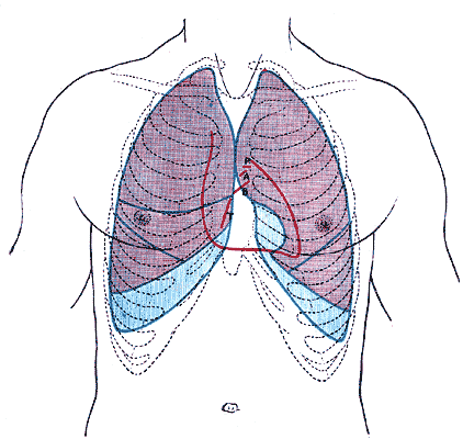
Surface anatomy
The opening and closing of the mitral valve is difficult to hear directly, but the flow of blood to the left ventricle is most audible at the apex of the heart, and so auscultation is usually performed at the intersection of the fifth intercostal space and the midclavicular line.
Additional images
-
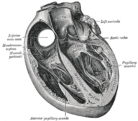 Section of the heart showing the ventricular septum. (Bicuspid valve visible at center.)
 -


Related Chapters
- Anatomy of the heart
- Pathophysiology
- Procedures to fix the mitral valve
Resources
- Template:SUNYAnatomyFigs - "Valves of the heart."
- Mitral Valve Repair at The Mount Sinai Hospital - "Mitral Valve Function"
- Surgical Anatomy of the Mitral Valve at echoincontext.com
- Cleveland Clinic Webchat - Latest Innovations in Mitral Valve Surgery with Dr. Marc Gillinov