Right ventricle
Editor-In-Chief: C. Michael Gibson, M.S., M.D. [1] Template:Infobox Anatomy
Overview
The right ventricle is one of four chambers (two atria and two ventricles) in the human heart. It receives de-oxygenated blood from the right atrium via the tricuspid valve, and pumps it into the pulmonary artery via the pulmonary valve.
It is triangular in form, and extends from the right atrium to near the apex of the heart.
Boundaries
Its anterosuperior surface is rounded and convex, and forms the larger part of the sternocostal surface of the heart.
Its under surface is flattened, rests upon the diaphragm, and forms a small part of the diaphragmatic surface of the heart.
Its posterior wall is formed by the ventricular septum, which bulges into the right ventricle, so that a transverse section of the cavity presents a semilunar outline.
Its upper and left angle forms a conical pouch, the conus arteriosus, from which the pulmonary artery arises.
A tendinous band, which may be named the tendon of the conus arteriosus, extends upward from the right atrioventricular fibrous ring and connects the posterior surface of the conus arteriosus to the aorta.
The wall of the right ventricle is thinner than that of the left, the proportion between them being as 1 to 3; it is thickest at the base, and gradually becomes thinner toward the apex.
The cavity equals in size that of the left ventricle, and is capable of containing about 85 c.c.
Components
Its interior presents the following parts for examination:
- right atrioventricular orifice
- opening of the pulmonary artery
- tricuspid valve
- pulmonary valve
- trabeculæ carneæ
- chordæ tendineæ
Additional images
-
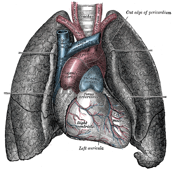 Front view of heart and lungs.
