Coronary venous anatomy
Editor-In-Chief: C. Michael Gibson, M.S., M.D. [1]
Associate Editor-In-Chief: Cafer Zorkun, M.D., Ph.D. [2]
Coronary Venography in CRT Implantation
After performing coronary venography the operator must then select the coronary vein tributary that will deliver the lead to a posterior and lateral location. Due to discrepancies between physicians in naming the branches of the coronary sinus and tributaries we rely on academic sources and journal reports for the correct nomenclature.
Description of Anatomy
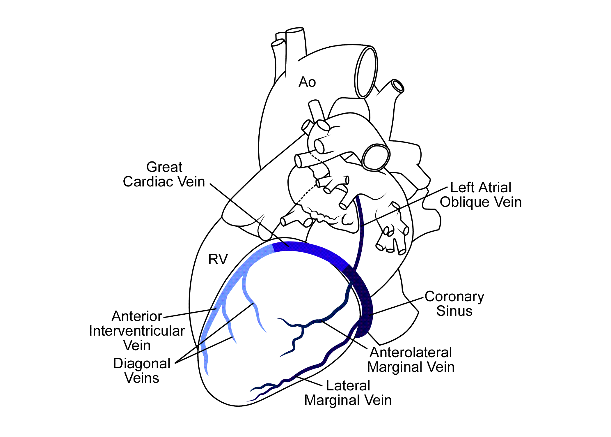
The coronary veins are described starting with the Anterior Interventicular Vein (AIV) which is defined as originating at the lower or middle third of the anterior interventricular groove. It connects with Diagonal Veins (DV) supplying the lateral and anterolateral portion of the left ventricle. It continues to run vertically and as it turns posterior at the AV groove and courses horizontally it becomes the Great Vein (GV). The Great Vein courses medial over the summit of the Left Ventricle.
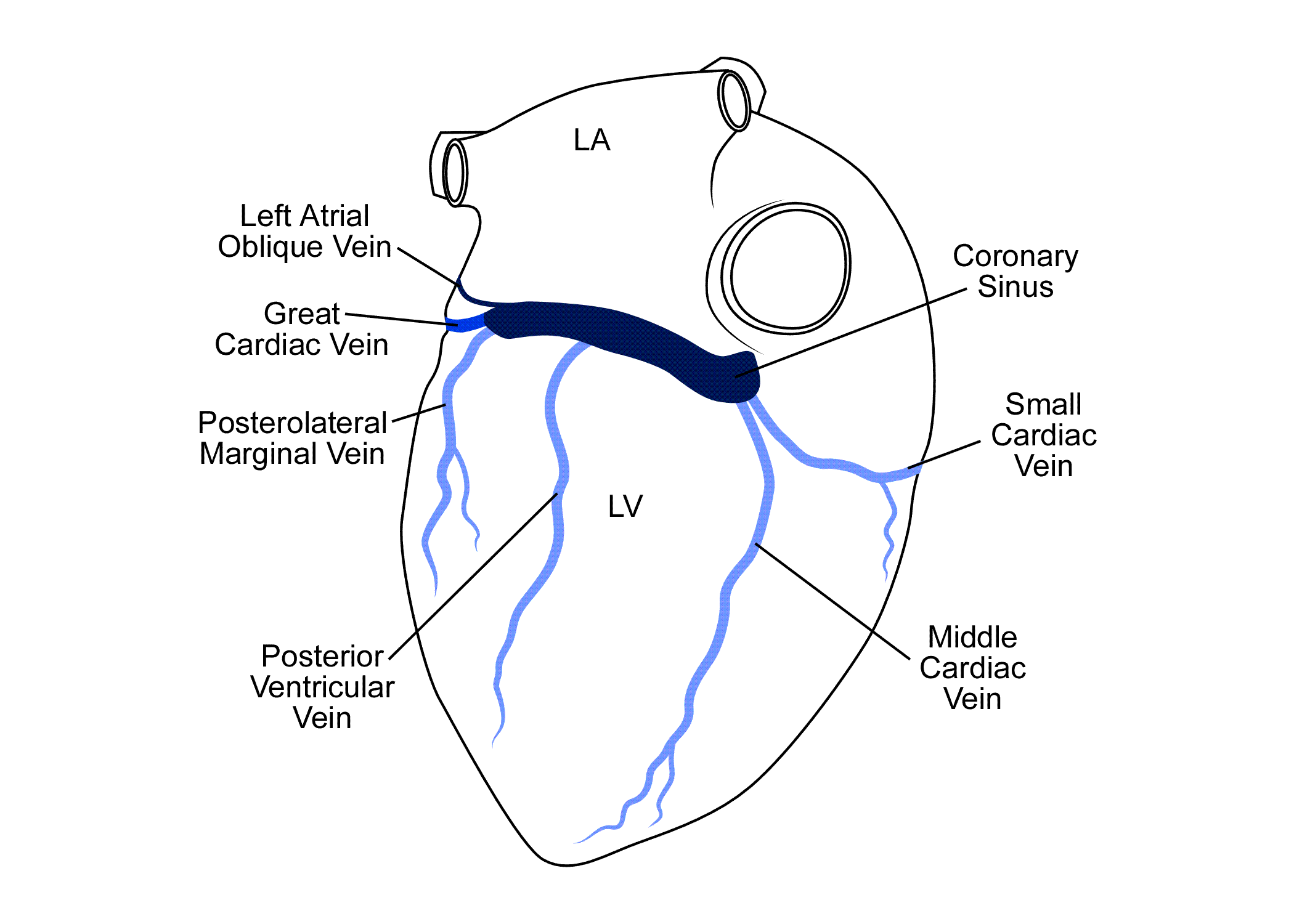
The Great Cardiac vein becomes the coronary sinus (CS) at the Left Atrial Oblique Vein of Marshall (LAOV). In cases were the LAOV is not present the Coronary Sinus begins at the valve of Vieussens.
The tributaries of the Coronary Sinus that originate off the lateral wall are referred to as Marginal Veins: These marginal veins are further defined by the location the veins empty into the Coronary Sinus in the LAO projection, such as the Anterolateral Marginal (AM), Lateral Marginal (LM), and Inferolateral Marginal (IM). Since posterior is not an accepted in echocardiographic standards Inferolateral Marginal is used in naming the CS tributary here for consistency, but Posterolateral Marginal may be used.
The Posterior Ventricular Vein (PVV) drains the inferior (posterior) aspect of the Left Ventricle. The Posterior Ventricular Vein is often confused with the Posterolateral marginal vein due to the similar location. The PVV is usually larger in caliber than the posterolateral marginal and sometimes drain into the Middle cardiac vein. The Middle Cardiac Vein (MCV) originates near the apex runs in the posterior interventricular groove and drains either directly into the rights atrium or into the Coronary Sinus just before it opens into the right atrium.
-
 Anterior View
 -
 Posterior View


Flouroscopic Views
Many cardiologists also prefer to use fluoroscopy for the identification of the location of the left ventricular lead tip after completion of the procedure. Since implantation of left ventricular leads occurs in the Electrophysiology lab with fluoroscopy we used fluoroscopic views to correspond with the short axis and long axis views of the heart. Using this method an accurate and consistent description of the location of the LV lead tip can be made at the time of implant and entered into the operative report rather than relying on the CXR for documentation of lead position. The RAO 30 projection is used to define the location of the left ventricle along the vertical axis of the heart dividing it into a basal, mid and apical third. The LAO 30 projection is used for the short axis views to define the circumferential location of the lead.
-
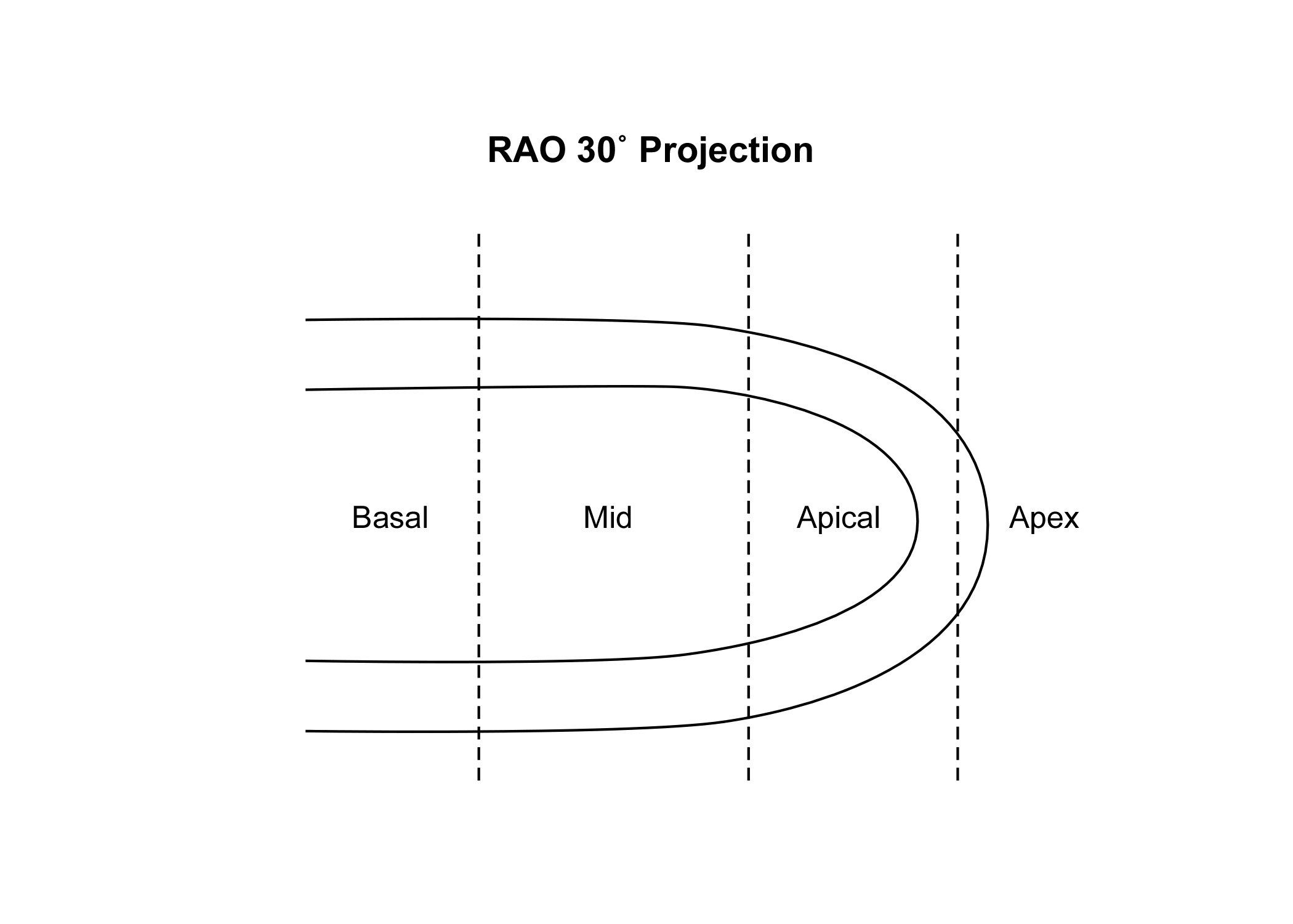 RAO 30 View
 -
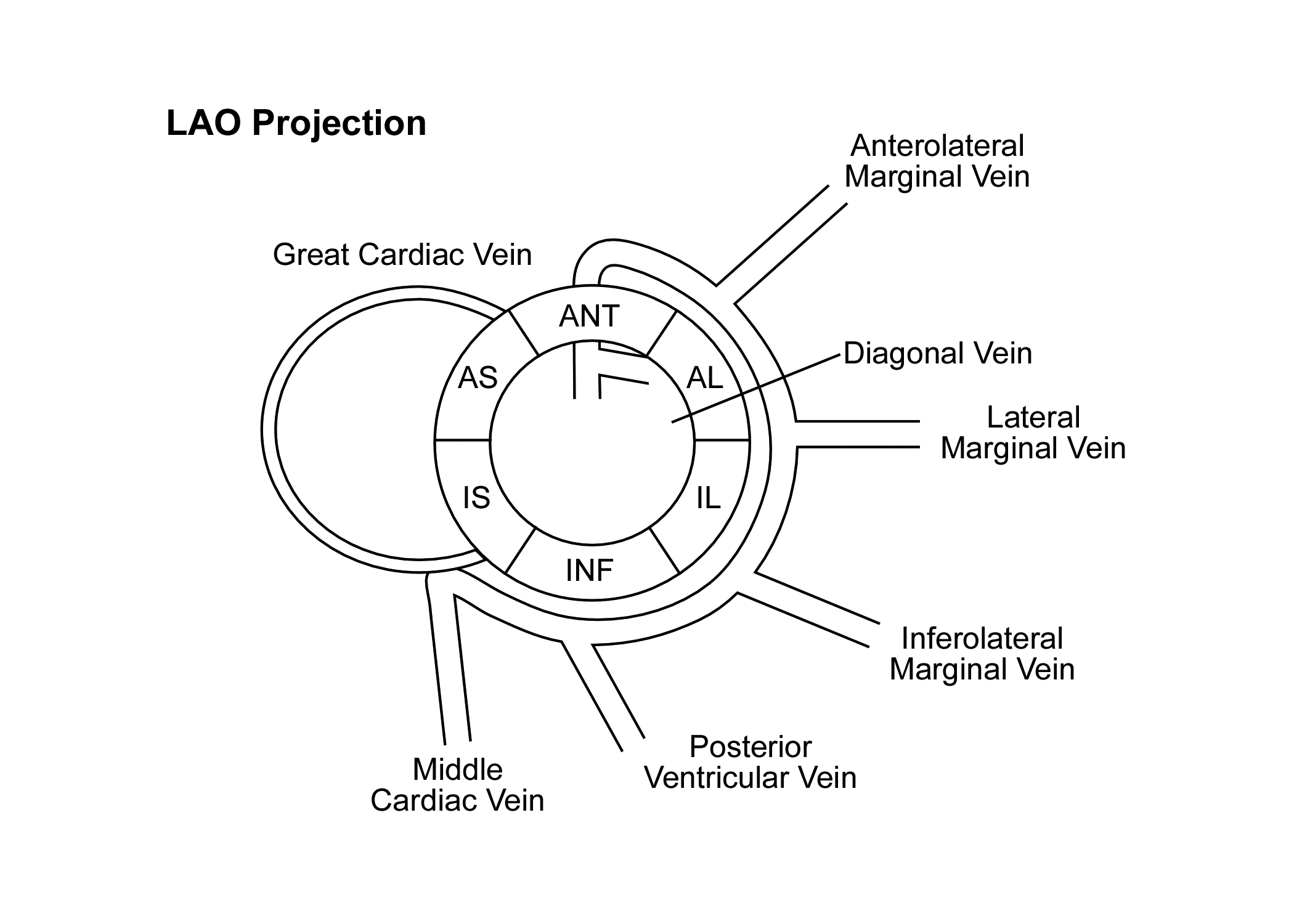 LAO 30 View

