Premature atrial contraction
| Premature atrial contraction | |
 | |
|---|---|
| 12 lead EKG shows Premature Atrial Contractions and bifascicular block (RBBB + LAFB) | |
| ICD-10 | I49.1 |
| ICD-9 | 427.61 |
| MeSH | D018880 |
Editor-In-Chief: C. Michael Gibson, M.S., M.D. [1]
Associate Editor-In-Chief: Cafer Zorkun, M.D., Ph.D. [2]
Overview
Premature atrial contractions or (PACs) are a type of premature heart beat, irregular heart beat or benign arrhythmia which start in the upper two chambers of the heart, also called atria. These aren't as serious as a Premature Ventricular Contraction and usually require no medical care. Individuals with the condition may report feeling that his or her heart "stops" after a symptom. PVCs are also called heart palpitations (although there are many other forms of arrhythmia).
Differential Diagnosis of Underlying Causes
- Alcohol
- Anxiety
- Caffeine
- Cocaine
- Coffeee
- Drugs
- Smoking
Diagnosis
Symptoms
There may be a sense of "a skipped beat" or "a thump in the chest" or neck. In many cases, the person feels nothing.
Signs
The pulse will feel irregular in a patient who has frequent PACs.
Electrocardiogram
The presence of PACs is diagnosed based upon either an EKG, Holter, or Cardiac Event Monitor.
- By definition the P waves are premature
- Morphology of the P' wave is different than the P wave in normal sinus rhythm. If its origin is close to that of the sinus node, then the P' morphology is hard to distinguish from the native sinus P wave.
- A PAC differs from a Premature Junctional Contraction (PJC) in that the PR interval is > 0.12 second in a PAC.
- The PR interval may be shorter than that in Normal Sinus Rhythm (NSR) if it is located closer to the AV node.
- The PR interval tends to lengthen when the coupling time to the PAC is short.
- A PAC may not be conducted to the ventricles and this is called a blocked PAC.
- The differential diagnosis in this scenario includes second degree AV block. In second degree AV block, the PP intervals remain constant.
- Usually the QRS is of normal duration, but occasionally there is aberrant conduction, most frequently of Right Bundle Branch Block (RBBB) morphology.
- Aberrancy is more likely to occur when the coupling time is shorter.
- Usually there is not a compensatory pause. The PAC resets the sinus node.
- Most these patients do not have organic heart disease.
- 64% of healthy subjects will have PACs on 24 hour Holter monitoring.
- Frequency higher in the elderly
- The QRS complex is normal
- There is normal T wave repolarization (not inverted to the other T waves)
Electrocardiogram Examples
-
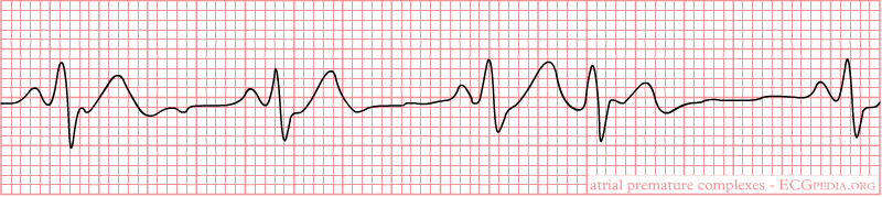
Premature Atrial Contractions
-
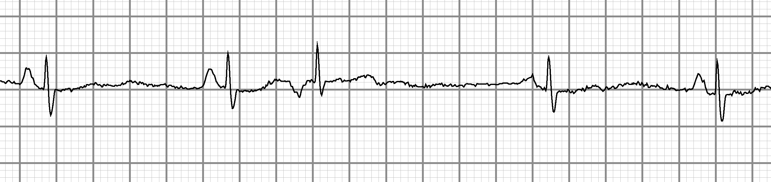
Premature Atrial Contractions
-
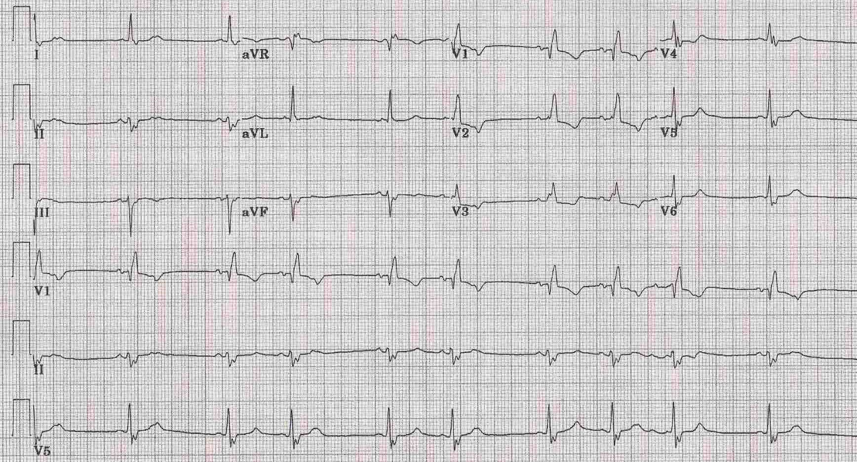
12 lead EKG shows Premature Atrial Contractions in patient with RBBB + LAFB
Prognosis
In general the prognosis of PACs is good, and is determined by the underlying condition that may trigger the PACs. In rare cases, a PAC can, like a PVC, trigger a more serious arrhythmia such as Atrial flutter or fibrillation. This is seldom seen in a healthy heart, but in a case that it does happen, medical attention should be sought to see what might have caused the problem and what steps can be taken to fix it. Unlike PVC's, PAC's generally do not compromise the hemodynamic system because the conduction throughout the AV node, and the ventricles of the heart are activated in a normal sequence.
Additional resources
- ECGpedia: Course for interpretation of ECG
- The whole ECG - A basic ECG primer
- 12-lead ECG library
- Simulation tool to demonstrate and study the relation between the electric activity of the heart and the ECG
- ECG information from Children's Hospital Heart Center, Seattle
- ECG Challenge from the ACC D2B Initiative
- National Heart, Lung, and Blood Institute, Diseases and Conditions Index
- A history of electrocardiography
- EKG Interpretations in infants and children