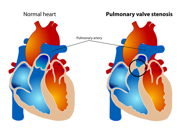
Pulmonary valve stenosis
| Pulmonary valve stenosis | |
 | |
|---|---|
| Pulmonary valve stenosis | |
| ICD-10 | I37.0, I37.2, Q22.1 |
| ICD-9 | 424.3, 746.02 |
| OMIM | 265500 |
| DiseasesDB | 11025 |
| MedlinePlus | 001096 |
| eMedicine | emerg/491 |
Editor-In-Chief: C. Michael Gibson, M.S., M.D. [1]
For an expanded discussion of this topic see right ventricular outflow tract obstruction
Overview
Pulmonary valve stenosis is a valvular heart disease in which outflow of blood from the right ventricle of the heart is obstructed at the level of the pulmonic valve. This results in the reduction of flow of blood to the lungs.
Demographics and Epidemiology
- Generally, pulmonary valve stenosis is a congenital defect narrowing of the pulmonary valve (the semilunar valve that separates the right ventricule from the pulmonary artery), but occasionally, it could also be presented in adults as a complication of another illness.
- It's one of the more common heart birth defects, and most cases are mild. If the pulmonary valve stenosis is moderate to severe, it will cause serious symptoms, requiring surgery which is highly successful.
- It occurs in about 1 of 10 children, and females are slightly more likely to be affected than males.
Etiology
- Congenital pulmonic stenosis is most common.
- Rheumatic involvement is rare, is usually part of multivalvular involvement, rarely leads to serious deformity.
- Carcinoid plaques can be present in the carcinoid syndrome. These result in constriction of the pulmonic valve ring, retraction and fusion of the valve cusps.
Anatomy
- Typically the valve is domed shaped with fused commissures.
- If the foramen ovale is patent, then right to left shunting can occur at the atrial level.
- If there is pulmonary atresia with an intact ventricular septum then these patients die soon after birth.
Diagnosis
Symptoms
Symptoms include cyanosis (usually visible in the nailbeds), and general symptoms of hypoxemia. When the stenosis is mild, it can go unnoticed for many years. If stenosis is severe, dizziness or frank syncope may develop, particularly with exercise.
Physical Examination
Neck
Echocardiography
2D echocardiography
- Thickened leaflets with systolic bowing in valvular stenosis.
- Difficult to distinguish between valvular, sub valvular and supra valvular stenosis with 2D echocardiography.
- Post stenotic pulmonary artery dilatation can be visualised sometimes.
Doppler echocardiography
- Ante grade velocity increased with corresponding maximum and mean pressure gradients.
- Pulmonary valve area can be calculated using the continuity equation.
- Pulmonary Valve Area = (Cross sectional areaRVOT * VTIRVOT)/ VTIPV
- The site of obstruction can be difficult to diagnose by 2D echo. Cautious use of colour flow mapping and PW Doppler can pin point the location of obstruction.
Severity Assessment
| Severity | mild | moderate | severe |
|---|---|---|---|
| Valve area | >1.0 | 1- 0.5 | <0.5 |
| Peak gradient (mm Hg) | <10-25 | 25-40 | >40 |
- Pulmonic Stenosis 1
<googlevideo>6761754875447006755&hl=en</googlevideo>
- Pulmonic Stenosis 2
<googlevideo>-5301172737736229119&hl=en</googlevideo>
- Pulmonic Stenosis 3
<googlevideo>-5141870933248575471&hl=en</googlevideo>
Treatment
Valve replacement or surgical repair (depending upon whether the stenosis is in the valve or vessel) may be indicated.
External links
- Overview at University of Maryland
- Overview at American Heart Association
- Pulmonary Stenosis information from Seattle Children's Hospital Heart Center