Supraventricular tachycardia
| Supraventricular tachycardia | |
 | |
|---|---|
| ICD-10 | I47.1 |
| ICD-9 | 427.89 |
| MeSH | D013617 |
Editor-In-Chief: C. Michael Gibson, M.S., M.D. [1]
Synonyms and keywords: SVT, paroxysmal supraventricular tachycardia, PSVT, paroxysmal atrial tachycardia; PAT; PAT with block
Overview
A supraventricular tachycardia (SVT) is a tachycardia or rapid rhythm of the heart in which the origin of the electrical signal is either the atria or the AV node. These rhythms, by definition, are either initiated or maintained by the atria or the AV node. This is in contrast to ventricular tachycardias, which are rapid rhythms that originate from the ventricles of the heart, that is, below the atria or AV node. The term SVT encompasses a large number of arrhythmias arising from the atria and AV node, and the term SVT is often incorrectly applied only to the subgroup of AV nodal re-entrant tachycardias.
Classification
SVTs can be classified in a number of ways. One way to classify them is via their electrophysiologic origin:
SVTs from a Sinoatrial Source
- Sinus tachycardia
- Inappropriate sinus tachycardia
- Sinoatrial node reentrant tachycardia (SANRT)
SVTs from an Atrial Source
- (Unifocal) Atrial tachycardia (AT)
- Multifocal atrial tachycardia (MAT)
- Atrial fibrillation with a rapid ventricular response
- Atrial flutter with a rapid ventricular response
SVTs from an Atrioventricular Source
- AV nodal reentrant tachycardia (AVNRT)
- AV reentrant tachycardia (AVRT)
- Junctional ectopic tachycardia (JET)
- Junctional tachycardia
Differentiating SVT from VT
Most supraventricular tachycardias have a narrow QRS complex on the EKG. It is not infrequent, however, for aberrant conduction to be be present, sometimes as a result of the more rapid rate of conduction. This widening of the QRS complex yields supraventricular tachycardia with aberrant conduction (SVTAC) which produces a wide-complex tachycardia that may mimic ventricular tachycardia (VT). In the clinical setting, it is important to determine whether a wide-complex tachycardia is an SVT or a ventricular tachycardia, since they are treated differently. Ventricular tachycardia has to be treated appropriately, since it can quickly degenerate to ventricular fibrillation and death. A number of different algorithms have been devised to determine whether a wide complex tachycardia is supraventricular or ventricular in origin.[1]
In general, a history of structural heart disease, ischemic heart disease or congestive heart failure increases the likelihood that the tachycardia is ventricular in origin.
Differentiating Among the Different Types of Supraventricular Tachycardia
The individual subtypes of SVT can be distinguished from each other by certain physiological and electrical characteristics, many of which present in the patient's EKG.
Supraventricular tachycardias must be differentiated from each other because the management strategies may vary:
Atrial Fibrillation
- Rate: 110 to 180 bpm
- Rhythm: Irregularly irregular
- P waves: Absent, fibrillatory waves
- PR interval: Absent
- QRS complex: Less than 0.12 seconds, consistent, and normal in morphology in the absence of abberant conduction
- Response to Maneuvers: Does not break with adenosine or vagal maneuvers
- Epidemiology and Demographics: More common in the elderly, following bypass surgery, in mitral valve disease, hyperthyroidism
Atrial Flutter
- Rate: 75 (4:1 block), 100 (3:1 block) and 150 (2:1 block) bpm, but 150 is most common
- Rhythm: Regular
- P waves: Sawtooth pattern of P waves at 250 to 350 beats per minute
- PR interval: Varies depending upon the magnitude of the block, but is short
- QRS complex: Less than 0.12 seconds, consistent, and normal in morphology
- Response to Maneuvers: Conduction may vary in response to drugs and maneuvers dropping the rate from 150 to 100 or to 75 bpm
- Epidemiology and Demographics: More common in the elderly, after alcohol
AV Nodal Reentry Tachycardia
- Rate: In adults the range is 140-250 bpm, but in children the rate can exceed 250 bpm.
- Rhythm: Regular
- P waves: The p wave is usually superimposed on or buried within the QRS complex
- PR interval: The PR interval cannot be calculated as the p wave is generally obscured by the QRS complex. In uncommon AVNRT, the p wave can appear after the QRS complex and before the T wave, and in atypical AVNRT, the p wave can appear just before the QRS complex.
- QRS complex: Less than 0.12 seconds, consistent, and normal in morphology in the absence of abberant conduction, QRS alternans may be present
- Response to Maneuvers: May break with adenosine or vagal maneuvers
- Epidemiology and Demographics: Accounts for 60%-70% of all SVTs. 80% to 90% of cases are due to antegrade conduction down a slow pathway and retrograde up a fast pathway.
AV Reciprocating Tachycardia
- Rate: More rapid than AVNRT
- Rhythm:
- P waves:
- PR interval:
- QRS complex: Less than 0.12 seconds, consistent, and normal in morphology
- Response to Maneuvers: May break with adenosine or vagal maneuvers
- Epidemiology and Demographics: More common in males, whereas AVNRT is more common in females, Occurs at a younger age
Junctional Tachycardia
- Rate: > 60 beats per minute
- Rhythm: Regular
- P waves: Usually inverted, may be burried in the QRS complex
- PR interval: The p wave is usually buried in the QRS complex
- QRS complex: Less than 0.12 seconds, consistent, and normal in morphology
- Response to Maneuvers: Does not break with adenosine or vagal maneuvers
- Epidemiology and Demographics: Common after heart surgery, digoxin toxicity, as an escape rhythm in AV block
Multifocal Atrial Tachycardia
- Rate: Atrial rate is > 100 beats per minute (bpm)
- Rhythm:
- P waves: P waves of varying morphology from at least three different foci, absence of one dominant atrial pacemaker, can be mistaken for atrial fibrillation if the P waves are of low amplitude
- PR interval: Variable PR intervals, RR intervals, and PP intervals
- QRS complex: Less than 0.12 seconds, consistent, and normal in morphology
- Response to Maneuvers: Does not terminate with adenosine or vagal maneuvers
- Epidemiology and Demographics: * High incidence in the elderly and in those with COPD
Sinus Node Reentry Tachycardia
- Rate:
- Rhythm:
- P waves: Upright P waves precede each regular, narrow QRS complex
- PR interval:
- QRS complex: Less than 0.12 seconds, consistent, and normal in morphology
- Response to Maneuvers: Although it cannot be distinguished on the surface 12 lead EKG from sinus tachycardia, SA node reentry tachycardia does often terminate with vagal maneuvers unlike sinus tachycardia.
- Epidemiology and Demographics:
Sinus tachycardia
- Rate: Greater than 100.
- Rhythm: Regular.
- P waves: Upright, consistent, and normal in morphology (if no atrial disease)
- PR interval: Between 0.12–0.20 seconds and shortens with increasing heart rate
- QRS complex: Less than 0.12 seconds, consistent, and normal in morphology
- Response to Maneuvers:
- Epidemiology and Demographics:
Ventricular Tachycardia
- Rate:
- Rhythm: Generally regular
- P waves: Normal morphology, upright, but dissociated from the QRS complex (i.e. "march through" the QRS complex)
- PR interval:
- QRS complex: Wide and greater than 0.12 seconds
- Response to Maneuvers: Does not terminate in response to adenosine or vagal maneuvers
- Epidemiology and Demographics:
- Risk Factors:: Occurs in the context of myocardial ischemia, myocardial infarction, congestive heart failure, drug toxicity, and inhereted channelopathies
Wolff-Parkinson-White syndrome
- Pathophysiology: Anatomically and functionally, the fast and slow pathways of AVNRT should not be confused with the accessory pathways that give rise to Wolff-Parkinson-White syndrome (WPW) syndrome or atrioventricular re-entrant tachycardia (AVRT). In AVNRT, the fast and slow pathways are located within the right atrium in close proximity to or within the AV node and exhibit electrophysiologic properties similar to AV nodal tissue. Accessory pathways that give rise to WPW syndrome and AVRT are located in the atrioventricular valvular rings, they provide a direct connection between the atria and ventricles, and have electrophysiologic properties similar to ventricular myocardium.
- Rate:
- Rhythm:
- P waves: In WPW with orthodromic conduction due to a bypass tract, the p wave generally follows the QRS complex, whereas in AVNRT, the p wave is generally buried in the QRS complex.
- PR interval:
- QRS complex: In WPW there is a delta wave and evidence of ventricular preexcitation if there is conduction to the ventrilce via antegrade conduction down an accessory pathway. It should be noted, however, that in some patients with WPW, a delta wave and pre-excitation may not be present because bypass tracts do not conduct antegrade.
- Response to Maneuvers: May break in response to procainamide, adenosine, vagal maneuvers
- Epidemiology and Demographics:
- Risk Factors: None, an inhereted disorder
Risk Factors
Underlying structural heart disease is generally absent. Often, there is no precipitant of an episode. Risk factors for precipitation of AVNRT include:
- Alcohol
- Anemia
- Anxiety
- Caffeine
- Chocolate
- Fever
- Hyperthyroidism
- Hypokalemia
- Hypomagnesemia
- Hypoxia
- Myocardial ischemia
- Menstruation
- Psychological stress
- Pulmonary embolism
- Stimulants
- Tea
- Theobromine in foods like tea, coffee and chocolate
- Theophylline
Natural History, Complications, Prognosis
SVTs may start and stop abruptly. Patients may develop syncope. The prognosis of an SVT is generally good in absence of underlying heart disease.
Natural History
The rhythm often ceases abruptly and spontaneously, particularly the most common form AVNRT. An episode generally last seconds to hours.
Complications
- Some patients will develop syncope during episodes of AVRNT. The mechanism of syncope may be due to a reduction of cardiac output and hemodynamic compromise as a result of the short ventricular filling time or alternatively it may be due to transient asystole due to tachycardia-mediated suppression of the sinus node when the rhythm terminates. Those patients who do become symptomatic during episodes of SVT (i.e. have syncope) should avoid activities where the occurrence of hemodynamic compromise would endanger their safety or that of others (like driving).
- In patients with underlying ischemic heart disease, demand-related myocardial ischemia, angina and even myocardial infarction and/or congestive heart failure can occur.
- Tachycardia mediated cardiomyopathy may develop if the SVT is chronic and does not terminate.
Prognosis
SVTs are rarely life threatening and in the absence of underlying structural heart disease, the prognosis is good. Radiofrequency ablation is curative in 95% of cases of AVNRT.
Diagnosis
Symptoms
Symptoms that are common to all types of SVT include the following:
- Anxiety
- Asystole may occur due to tachycardia-mediated suppression of the sinus node when the rhythm in AVNRT
- Chest pain or sensation of tightness
- Dizziness, or lightheadedness (near-faint), or fainting
- Lightheadedness
- Palpitations - The sensation of the heart racing, fluttering or pounding strongly in the chest or the carotid arteries
- Shortness of breath
- Syncope in cases of AVNRT
- Sweating
- Tachycardia mediated cardiomyopathy may develop if the AVNRT is chronic and does not terminate.
Diagnosis
-
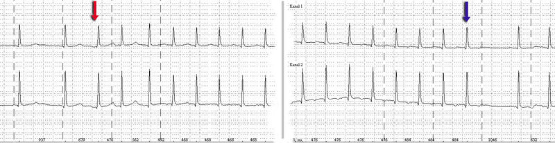
Holter monitor-Imaging with start (red arrow) and end (blue arrow) of a SV-tachycardia with a pulse frequency of about 128/min.
- Sinus tachycardia is considered "appropriate" when a reasonable stimulus, such as the catecholamine surge associated with fright, stress, or physical activity, provokes the tachycardia. It is distinguished by a presentation identical to a normal sinus rhythm except for its fast rate (>100 beats per minute in adults).
- Sinoatrial node reentrant tachycardia (SANRT) is caused by a reentry circuit localised to the SA node, resulting in a normal-morphology p-wave that falls before a regular, narrow QRS complex. It is therefore impossible to distinguish on the EKG from ordinary sinus tachycardia. It may however be distinguished by its prompt response to Vagal manouvres.
- (Unifocal) Atrial tachycardia is tachycardia resultant from one ectopic foci within the atria, distinguished by a consistent p-wave of abnormal morphology that fall before a narrow, regular QRS complex.
- Multifocal atrial tachycardia (MAT) is tachycardia resultant from at least three ectopic foci within the atria, distinguished by p-waves of at least three different morphologies that all fall before regular, narrow QRS complexes.
- Atrial fibrillation is not, in itself, a tachycardia, but when it is associated with a rapid ventricular response greater than 100 beats per minute, it becomes a tachycardia. A-fib is characteristically an "irregularly irregular rhythm" both in its atrial and ventricular depolarizations. It is distinguished by fibrillatory p-waves that, at some point in their chaos, stimulate a response from the ventricles in the form of irregular, narrow QRS complexes.
- Atrial flutter, is caused by a re-entry rhythm in the atria, with a regular rate of about 300 beats per minute. On the EKG, this appears as a line of "sawtooth" p-waves. The AV node will not usually conduct such a fast rate, and so the P:QRS usually involves a 2:1 or 4:1 block pattern, (though rarely 3:1, and most rarely and sometimes fatally 1:1). Because the ratio of P to QRS is usually consistent, A-flutter is often regular in comparison to its irregular counterpart, A-fib. Atrial Flutter is also not necessarily a tachycardia unless the AV node permits a ventricular response greater than 100 beats per minute.
- AV nodal reentrant tachycardia (AVNRT) is also sometimes referred to as a junctional reciprocating tachycardia. It involves a reentry circuit forming just next to or within the AV node itself. The circuit most often involves two tiny pathways one faster than the other, within the AV node. Because the AV node is immediately between the atria and the ventricle, the re-entry circuit often stimulates both, meaning that a retrogradely conducted p-wave is buried within or occurs just after the regular, narrow QRS complexes.
- Atrioventricular reentrant tachycardia (AVRT) also results from a reentry circuit, although one physically much larger than AVNRT. One portion of the circuit is usually the AV node, and the other, an abnormal accessory pathway from the atria to the ventricle. Wolff-Parkinson-White syndrome is a relatively common abnormality with an accessory pathway, the Bundle of Kent crossing the A-V valvular ring.
- In orthodromic AVRT, atrial impulses are conducted down through the AV node and retrogradely re-enter the atrium via the accessory pathway. A distinguishing characteristic of orthodromic AVRT can therefore be a p-wave that follows each of its regular, narrow QRS complexes, due to retrograde conduction.
- In antidromic AVRT, atrial impulses are conducted down through the accessory pathway and re-enter the atrium retrogradely via the AV node. Because the accessory pathway initiates conduction in the ventricles ouside of the bundle of His, the QRS complex in antidromic AVRT is often wider than usual, with a delta wave.
- Finally, Junctional Ectopic Tachycardia or JET is a rare tachycardia caused by increased automaticity of the AV node itself initiating frequent heart beats. On the EKG, junctional tachycardia often presents with abnormal morphology p-waves that may fall anywhere in relation to a regular, narrow QRS complex.
Acute Treatment
In general, SVT is not life threatening, but episodes should be treated or prevented. While some treatment modalities can be applied to all SVTs with impunity, there are specific therapies available to cure some of the different sub-types. Cure requires intimate knowledge of how and where the arrhythmia is initiated and propagated.
The SVTs can be separated into two groups, based on whether they involve the AV node for impulse maintenance or not. Those that involve the AV node can be terminated by slowing conduction through the AV node. Those that do not involve the AV node will not usually be stopped by AV nodal blocking manoevres. These manoevres are still useful however, as transient AV block will often unmask the underlying rhythm abnormality.
AV nodal blocking can be achieved in at least three different ways:
Physical maneuvers
A number of physical maneuvers cause increased AV nodal block, principally through activation of the parasympathetic nervous system, conducted to the heart by the Vagus nerve. These manipulations are therefore collectively referred to as vagal maneuver.
The best recognised of these is the Valsalva maneuver, which increases intra-thoracic pressure and affects baro-receptors (pressure sensors) within the arch of the aorta. This can be achieved by asking the patient to hold their breath and "bear down" as if straining to pass a bowel motion, or less embarrassingly, by getting them to hold their nose and blow out against it. Plunging the face into, or just drinking a glass of ice cold water is also often effective. Firmly pressing the bulb at the top of one of the carotid arteries in the neck (carotis sinus massage, stimulating carotid baro-receptors) is also effective, but not recommended for those without adequate medical training.
Drug Treatment
Another modality involves treatment with medications. Prehospital care providers and hospital clinicians might administer Adenosine, an ultra short acting AV nodal blocking agent. If this works, followup therapy with Diltiazem, Verapamil or Metoprolol may be indicated. SVT that does NOT involve the AV node may respond to other anti-arrhythmic drugs such as Sotalol or Amiodarone.
In pregnancy, Metoprolol is the treatment of choice as recommended by the American Heart Association.
Electrical Cardioversion
If physical maneuvers or drugs do not work, or if the patient is extremely unstable, a DC shock delivered to the chest (synchronized cardioversion) may also be used, and is almost always effective.
Prevention & Cure
Once the acute episode has been terminated, ongoing treatment may be indicated to prevent a recurrence of the arrhythmia. Patients who have a single isolated episode, or infrequent and minimally symptomatic episodes usually do not warrant any treatment except observation.
Patients who have more frequent or disabling symptoms from their episodes generally warrant some form of preventative therapy. A variety of drugs including simple AV nodal blocking agents like beta-blockers and verapamil, as well as anti-arrhythmics may be used, usually with good effect, although the risks of these therapies need to be weighed against the potential benefits.
For supraventricular tachycardia caused by a re-entrant pathway, another form of treatment is radiofrequency ablation. This is a low risk procedure that uses a catheter inside the heart to deliver radiofrequency energy to locate and destroy the abnormal electrical pathways. Ablation has been shown to be highly effective: up to 99% effective in eliminating AVNRT, and similar results in typical Atrial flutter.
Paroxysmal atrial tachycardia is a period of very rapid and regular heart beats that begins and ends abruptly. The heart rate is usually between 160 and 200 beats per minute. This condition is also known as paroxysmal supraventricular tachycardia.
See also
- Tachycardia
- AV nodal reentrant tachycardia (AVNRT)
- AV reentrant tachycardia (AVRT)
- Inappropriate Sinus Tachycardia
- Ashman phenomenon