Aortic dissection MRI
|
Aortic dissection Microchapters |
|
Diagnosis |
|---|
|
Treatment |
|
Special Scenarios |
|
Case Studies |
|
|
Editor-In-Chief: C. Michael Gibson, M.S., M.D. [1]
Associate Editor-In-Chief: Cafer Zorkun, M.D., Ph.D. [2]
MRI
Magnetic resonance imaging (MRI) is currently the gold standard test for the detection and assessment of aortic dissection, with a sensitivity of 98% and a specificity of 98%. An MRI examination of the aorta will produce a three-dimensional reconstruction of the aorta, allowing the physician to determine the location of the intimal tear, the involvement of branch vessels, and locate any secondary tears. It is a non-invasive test, does not require the use of iodinated contrast material, and can detect and quantitate the degree of aortic insufficiency.
The disadvantage of the MRI scan in the face of aortic dissection is that it has limited availability and is often located only in the larger hospitals, and the scan is relatively time consuming. Due to the high intensity of the magnetic waves used during MRI, an MRI scan is contraindicated in individuals with metallic implants. In addition, many individuals succumb to claustrophobia while in the MRI scanning tube.
-
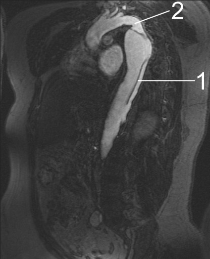
MRI of an aortic dissection. 1 Aorta descendens with dissection. 2 Aortic isthmus.

References
Acknowledgements
The content on this page was first contributed by: David Feller-Kopman, MD and C. Michael Gibson M.S., M.D.