Ventricular arrhythmias: Difference between revisions
| Line 288: | Line 288: | ||
=== Electrophysiological Testing in Patients With Syncope Recommendations === | === Electrophysiological Testing in Patients With Syncope Recommendations === | ||
=== | {|class="wikitable" | ||
|- | |||
| colspan="1" style="text-align:center; background:LightGreen"|[[ACC AHA Guidelines Classification Scheme#Classification of Recommendations|Class I]] | |||
|- | |||
| bgcolor="LightGreen"| <nowiki>"</nowiki>'''1''' EP testing is recommended in patients with syncope of unknown cause with impaired LV function or structural heart disease. ''([[ACC AHA Guidelines Classification Scheme#Level of Evidence|Level of Evidence: B]])'' <nowiki>"</nowiki> | |||
|} | |||
{|class="wikitable" | |||
|- | |||
| colspan="1" style="text-align:center; background:LemonChiffon"|[[ACC AHA Guidelines Classification Scheme#Classification of Recommendations|Class IIa]] | |||
|- | |||
|bgcolor="LemonChiffon"| <nowiki>"</nowiki>'''1''' EP testing can be useful in patients with syncope when bradyarrhythmias or tachyarrhythmias are suspected and in whom noninvasive diagnostic studies are not conclusive. ''([[ACC AHA Guidelines Classification Scheme#Level of Evidence|Level of Evidence: B]])''<nowiki>"</nowiki> | |||
|} | |||
=== Ablation Recommendations === | === Ablation Recommendations === | ||
Revision as of 16:02, 3 October 2012
Editor-In-Chief: C. Michael Gibson, M.S., M.D. [1]; Associate Editor: Cafer Zorkun, M.D., Ph.D. [2]
Causes of Ventricular Arrhythmias
Cardiac causes
Acute and chronic ischemic heart disease
Noncardiac causes
Stimulants: caffeine, cocaine, alcohol
Metabolic abnormalities: acidosis, hypoxemia, hyperkalemia, hypokalemia, hypomagnesemia
Drugs: digoxin (Lanoxin), theophylline, antipsychotics, tricyclic antidepressants, antiarrhythmics with proarrhythmic potential [e.g., flecainide (Tambocor), dofetilide (Tikosyn), sotalol (Betapace) and quinidine)
| example | regularity | atrial frequency | ventricular frequency | origin (SVT/VT) | p-wave | effect of adenosine | |
|---|---|---|---|---|---|---|---|
| Wide complex (QRS>0.12) | |||||||
| Ventricular Tachycardia | regular (mostly) | 60-100 bpm | 110-250 bpm | ventricle (VT) | AV-dissociation | no rate reduction (sometimes accelerates) | |
| Ventricular Fibrillation | irregular | 60-100 bpm | 400-600 bpm | ventricle (VT) | AV-dissociation | none | |
| Ventricular Flutter | regular | 60-100 bpm | 150-300 bpm | ventricle (VT) | AV-dissociation | none | |
| Accelerated Idioventricular Rhythm | regular (mostly) | 60-100 bpm | 50-110 bpm | ventricle (VT) | AV-dissociation | no rate reduction (sometimes accelerates) | |
| Torsade de Pointes | regular | 150-300 bpm | ventricle (VT) | AV-dissociation | no rate reduction (sometimes accelerates) | ||
| Bundle-branch re-entrant tachycardia* | regular | 60-100 bpm | 150-300 bpm | ventricles (VT) | AV-dissociation | no rate reduction | |
| (*) Bundle-branch re-entrant tachycardia is extremely rare | |||||||
Premature Ventricular Complexes (PVCs)
A premature ventricular complex is characterized by the premature occurrence of a QRS complex that is bizarre in shape and lasts longer than 120 msec. The T wave is large and usually of opposite polarity to the QRS complex.
A premature ventricular complex is usually followed by a full compensatory pause. The term “ventricular bigeminy” refers to alternating normal sinus and premature ventricular complexes. Three or more successive premature ventricular complexes are arbitrarily defined as ventricular tachycardia. Premature ventricular complexes become more prevalent with increasing age and occur in association with a variety of stimuli.
It is important to determine whether underlying structural heart disease is present and left ventricular function is impaired.Other common causes include electrolyte abnormalities, stimulants, and some medications.
Management
Patients Without Heart Disease
In the absence of heart disease, PVCs are associated with little or no increased risk of developing a dangerous arrhythmia. In this situation, the risk-to-benefit ratio of antiarrhythmic drug therapy does not support routine treatment.[1] It is important to review medications, determine if stimulants are being used, and correct electrolyte abnormalities. If no underlying cause is found, the optimal approach is patient reassurance. Patients should be made aware of the potential dangers of antiarrhythmic drug therapy as determined in the Cardiac Arrhythmia Suppression Trials (CAST and CAST II).[2] [3]
CAST showed that the risk of dying increased, rather than decreased, with successful long-term suppression of premature ventricular complexes after myocardial infarction in older patients. At best, CAST II showed no impact on long-term survival from drug treatment that successfully suppressed premature ventricular complexes. If patients with multiple premature ventricular complexes have severe, disabling symptoms, beta blockers are the safest initial choice.Referral to a cardiologist is indicated if beta-blocker therapy is not effective. In this situation, the next agents to be tried would be class I antiarrhythmic drugs, such as flecainide (Tambocor) and amiodarone (Cordarone), although radiofrequency ablation of an ectopic focus may also be an appropriate treatment.
Patients with Structural Heart Disease
The occurrence of premature ventricular complexes in patients with structural heart disease has been shown to significantly increase the risk of subsequent morbidity and mortality. Coronary heart disease, cardiomyopathy, and congestive heart failure are the major cardiac diseases associated with unfavorable outcomes in patients with premature ventricular complexes.
Ventricular Tachycardia
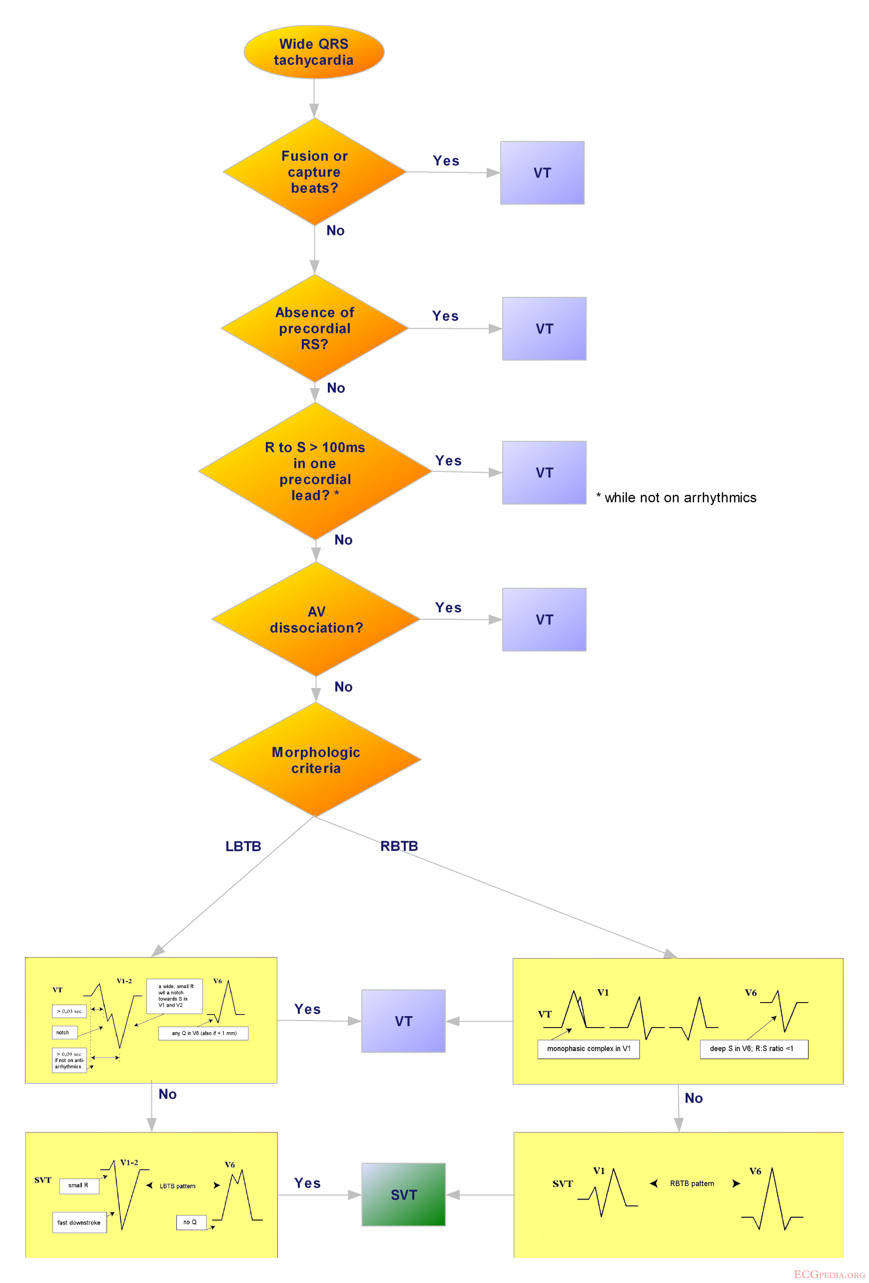
Ventricular tachycardia refers to a rhythm originating from a ventricular ectopic focus at a rate >100 bpm. The electrocardiogram shows a wide complex tachycardia with no associated P waves.
In patients with bundle branch block,Wolff-Parkinson-White syndrome, or aberrant conduction, supraventricular tachycardia can resemble ventricular tachycardia. Because of the morbidity and mortality associated with untreated ventricular tachycardia, any wide-complex tachycardia should be assumed to be ventricular tachycardia until proved otherwise. Physicians should keep in mind that patients with ventricular tachycardia can have minimal symptoms.
Management of Ventricular Tachycardia
The mortality rate within two years is reported to be higher than 30% in patients with non sustained ventricular tachycardia on Holter monitoring and impaired left ventricular function.[4] Two large multicenter trials [5] [6] showed a clear advantage for automatic cardioverter defibrillator implantation over drug therapy in patients who had a malignant ventricular arrhythmia or who had been resuscitated from sudden cardiac death. The selection of high-risk patients for defibrillator implantation should be based on left ventricular function and the findings of electrophysiologic studies.
Implantable defibrillators appear to be most beneficial in patients with a low ejection fraction who are noted to have frequent premature ventricular complexes, non sustained ventricular tachycardia on Holter monitoring, and a history of syncope or nearsyncope. It is critical to rule out coronary heart disease and to optimize the treatment of congestive heart failure in these patients. When ventricular tachycardia is diagnosed in relatively asymptomatic patients, medical treatment should be attempted. New recommendations from the American Heart Association [7] emphasize the initial use of 300 mg of iv. administered amiodarone, followed by repeated 150 mg iv. doses every 8-10 minutes, in patients with pulseless VT.
Patients with stable ventricular tachycardia should be given 150 mg of amiodarone intravenously over 10 minutes, followed by an infusion at 1 mg/minute for 6 hours and then at 0.5 mg/minute until the VT converts to sinus rhythm or another less dangerous rhythm. The alternative treatment is intravenously administered lidocaine (Xylocaine), given first in a 100 mg bolus (or 1 mg/kg) and then in an infusion at 1-4 mg/minute.
In hemodynamically unstable patients, electrical cardioversion should be attempted in accordance with the recently revised advanced cardiac life support (ACLS) protocols.
Automatic implantable cardioverter defibrillators (ICD) are considered the most effective treatment for patients with life-threatening VT or VF. According to expert recommendations, implantation of an automatic cardioverter defibrillator should be considered in these situations[8]:
1. Cardiac arrest resulting from ventricular fibrillation or tachycardia not caused by a transient or reversible cause
2. Spontaneous, sustained ventricular tachycardia
3. Syncope of undetermined origin and sustained VT or ventricular fibrillation (VF) induced during electrophysiologic studies
4. Non sustained VT with coronary artery disease and LV dysfunction if VT or VF is induced during electrophysiologic studies.
ACC/AHA/ESC 2006 Guidelines for Management of Patients With Ventricular Arrhythmias (DO NOT EDIT) [9]
Resting Electrocardiogram Recommendations
| Class I |
| "* Resting 12-lead ECG is indicated in all patients who are evaluated for ventricular arrhythmias. (Level of Evidence: A). " |
Exercise Testing Recommendations
| Class I |
| "1 Exercise testing is recommended in adult patients with ventricular arrhythmias who have an intermediate or greater probability of having CHD by age, gender, and symptoms* to provoke ischemic changes or ventricular arrhythmias. (Level of Evidence: B) *See Table 4 in the ACC/AHA 2002 Guideline Update for Exercise Testing (141) for further explanation of CHD probability." |
| "2 Exercise testing, regardless of age, is useful in patients with known or suspected exercise-induced ventricular arrhythmias, including catecholaminergic VT, to provoke the arrhythmia, achieve a diagnosis, and determine the patient’s response to tachycardia. (Level of Evidence: B)" |
| Class III |
| "1 See Table 1 in the ACC/AHA 2002 Guideline Update for Exercise Testing (141) for contraindications. (Level of Evidence: C)" |
| Class IIa |
| "1 Exercise testing can be useful in evaluating response to medical or ablation therapy in patients with known exercise-induced ventricular arrhythmias. (Level of Evidence: B)" |
| Class IIb |
| "1 Exercise testing may be useful in patients with ventricular arrhythmias and a low probability of CHD by age, gender, and symptoms.* (Level of Evidence: C) *See Table 4 in the ACC/AHA 2002 Guideline Update for Exercise Testing (141) for further explanation of CHD probability." |
| "2 Exercise testing may be useful in the investigation of isolated premature ventricular complexes (PVCs) in middle-aged or older patients without other evidence of CHD. (Level of Evidence: C)" |
Ambulatory Electrocardiography Recommendations
| Class I |
| "1 Ambulatory ECG is indicated when there is a need to clarify the diagnosis by detecting arrhythmias, QTinterval changes, T-wave alternans (TWA), or ST changes, to evaluate risk, or to judge therapy. (Level of Evidence: A) " |
| "2 Event monitors are indicated when symptoms are sporadic to establish whether or not they are caused by transient arrhythmias. (Level of Evidence: B) " |
| "3 Implantable recorders are useful in patients with sporadic symptoms suspected to be related to arrhythmias such as syncope when a symptom-rhythm correlation cannot be established by conventional diagnostic techniques. (Level of Evidence: B) " |
Electrocardiographic Techniques and Measurements Recommendations
| Class IIa
"1 It is reasonable to use TWA to improve the diagnosis and risk stratification of patients with ventricular arrhythmias or who are at risk for developing lifethreatening ventricular arrhythmias. (Level of Evidence: A) " |
| Class IIb
"1 ECG techniques such as signal-averaged ECG (SAECG), heart rate variability (HRV), baroflex sensitivity, and heart rate turbulence may be useful to improve the diagnosis and risk stratification of patients with ventricular arrhythmias or who are at risk of developing life-threatening ventricular arrhythmias. (Level of Evidence: B) " |
Left Ventricular Function and Imaging Recommendations
| Class I |
| "1 Echocardiography is recommended in patients with ventricular arrhythmias who are suspected of having structural heart disease. (Level of Evidence: B) " |
| "2 Echocardiography is recommended for the subset of patients at high risk for the development of serious ventricular arrhythmias or SCD, such as those with dilated, hypertrophic, or RV cardiomyopathies, AMI survivors, or relatives of patients with inherited disorders associated with SCD. (Level of Evidence: B) " |
| "3 Exercise testing with an imaging modality (echocardiography or nuclear perfusion [single-photon emission computed tomography (SPECT)]) is recommended to detect silent ischemia in patients with ventricular arrhythmias who have an intermediate probability of having CHD by age, symptoms, and gender and in whom ECG assessment is less reliable because of digoxin use, LVH, greater than 1-mm ST-segment depression at rest, WPW syndrome, or LBBB. (Level of Evidence: B) " |
| "4 Pharmacological stress testing with an imaging modality (echocardiography or myocardial perfusion SPECT) is recommended to detect silent ischemia in patients with ventricular arrhythmias who have an intermediate probability of having CHD by age, symptoms, and gender and are physically unable to perform a symptomlimited exercise test. (Level of Evidence: B) " |
| Class IIa |
| "1 MRI, cardiac computed tomography (CT), or radionuclide angiography can be useful in patients with ventricular arrhythmias when echocardiography does not provide accurate assessment of LV and RV function and/or evaluation of structural changes. (Level of Evidence: B)" |
| "2 Coronary angiography can be useful in establishing or excluding the presence of significant obstructive CHD in patients with life-threatening ventricular arrhythmias or in survivors of SCD, who have an intermediate or greater probability of having CHD by age, symptoms, and gender. (Level of Evidence: C)" |
| "3 LF imaging can be useful in patients undergoing biventricular pacing. (Level of Evidence: C) " |
Electrophysiological Testing in Patients With Coronary Heart Disease Recommendations
| Class I |
| "1 EP testing is recommended for diagnostic evaluation of patients with remote MI with symptoms suggestive of ventricular tachyarrhythmias, including palpitations, presyncope, and syncope. (Level of Evidence: B) " |
| "2 EP testing is recommended in patients with CHD to guide and assess the efficacy of VT ablation. (Level of Evidence: B) " |
| "3 EP testing is useful in patients with CHD for the diagnostic evaluation of wide-QRS-complex tachycardias of unclear mechanism. (Level of Evidence: C) " |
| Class IIa |
| "1 EP testing is reasonable for risk stratification in patients with remote MI, NSVT, and LVEF equal to or less than 40%. (Level of Evidence: B)" |
Electrophysiological Testing in Patients With Syncope Recommendations
| Class I |
| "1 EP testing is recommended in patients with syncope of unknown cause with impaired LV function or structural heart disease. (Level of Evidence: B) " |
| Class IIa |
| "1 EP testing can be useful in patients with syncope when bradyarrhythmias or tachyarrhythmias are suspected and in whom noninvasive diagnostic studies are not conclusive. (Level of Evidence: B)" |
Ablation Recommendations
| “ |
Class I
Class III
Class IIa
Class IIb
|
” |
Management of Cardiac Arrest Recommendations
| “ |
Class I
Class IIa
Class IIb
|
” |
Ventricular Tachycardia Associated With Low Troponin Myocardial Infarction Recommendations
| “ |
Class I
|
” |
Sustained Monomorphic Ventricular Tachycardia Recommendations
| “ |
Class I
Class III
Class IIa
Class IIb
|
” |
Repetitive Monomorphic Ventricular Tachycardia Recommendations
| “ |
Class IIaIntravenous amiodarone, beta blockers, and intravenous procainamide (or sotalol or ajmaline in Europe) can be useful for treating repetitive monomorphic VT in the context of coronary disease (375) and idiopathic VT. (Level of Evidence: C) |
” |
Polymorphic VT Recommendations
| “ |
Class I
Class IIbIntravenous lidocaine may be reasonable for treatment of polymorphic VT specifically associated with acute myocardial ischemia or infarction. (Level of Evidence: C) |
” |
Torsades de Pointes Recommendations
| “ |
Class I
Class IIa
Class IIb
|
” |
Incessant Ventricular Tachycardia Recommendations
| “ |
Class I
Class IIa
Class IIb
|
” |
Left Ventricular Dysfunction Due to Prior Myocardial Infarction Recommendations
| “ |
Class I
Class IIa
|
” |
Resources
1. Zipes DP, Camm AJ, Borggrefe M, et al., ACC/AHA/ESC 2006 Guidelines for Management of Patients With Ventricular Arrhythmias and the Prevention of Sudden Cardiac Death A Report of the American College of Cardiology/American Heart Association Task Force and the European Society of Cardiology Committee for Practice Guidelines (Writing Committee to Develop Guidelines for Management of Patients With Ventricular Arrhythmias and the Prevention of Sudden Cardiac Death)Circulation 2006;114;e385-e484
2. Kesh Hebbar A, Hueston WJ, Management of Common Arrhythmias: Part II.Ventricular Arrhythmias and Arrhythmias in Special Populations Am Fam Physician 2002;65:2491-6.
References
- ↑ Kennedy HL, Whitlock JA, Sprague MK, Kennedy LJ, Buckingham TA, Goldberg RJ. Long-term follow-up of asymptomatic healthy subjects with frequent and complex ventricular ectopy. N Engl J Med 1985;312:193-7.
- ↑ Echt DS, Liebson PR, Mitchell LB, Peters RW, Obias-Manno D, Barker AH, et al. Mortality and morbidity in patients receiving encainide, flecainide, or placebo. The Cardiac Arrhythmia Suppression Trial. N Engl J Med 1991;324:781-8
- ↑ Effect of the antiarrhythmic agent moricizine on survival after myocardial infarction. The Cardiac Arrhythmia Suppression Trial II Investigators. N Engl J Med 1992;327:227-33.
- ↑ Wilber DJ, Garan H, Finkelstein D, Kelly E, Newell J, McGovern B, et al. Out-of-hospital cardiac arrest. Use of electrophysiologic testing in the prediction of long-term outcome. N Engl J Med 1988;318:19-24.
- ↑ Moss AJ, Hall WJ, Cannom DS, Daubert JP, Higgins SL, Klein H, et al. Improved survival with an implanted defibrillator in patients with coronary disease at high risk for ventricular arrhythmia. Multicenter Automatic Defibrillator Implantation Trial Investigators. N Engl J Med 1996;335:1933-40.
- ↑ A comparison of antiarrhythmic drug therapy with implantable defibrillators in patients resuscitated from near-fatal ventricular arrhythmias. The Antiarrhythmics versus Implantable Defibrillators (AVID) Investigators. N Engl J Med 1997;337:1576-83.
- ↑ Gazmuri RJ, Nadkarni VM, Nolan JP et al., Scientific Knowledge Gaps and Clinical Research Priorities for Cardiopulmonary Resuscitation and Emergency Cardiovascular Care Identified During the 2005 International Consensus Conference on E and CPR Science With Treatment Recommendations A Consensus Statement From the International Liaison Committee on Resuscitation (American Heart Association, Australian Resuscitation Council, European Resuscitation Council, Heart and Stroke Foundation of Canada, Inter American Heart Foundation, Resuscitation Council of Southern Africa, and the New Zealand Resuscitation Council); the American Heart Association Emergency Cardiovascular Care Committee; the Stroke Council; and the Cardiovascular Nursing Council. Circulation 2007 http://circ.ahajournals.org/cgi/content/full/116/21/2501
- ↑ Gregoratos G, Cheitlin MD, Conill A, Epstein AE, Fellows C, Ferguson TB Jr, et al. ACC/AHA guidelines for implantation of cardiac pacemakers and antiarrhythmia devices: executive summary: a report of the American College of Cardiology/American Heart Association Task Force on Practice Guidelines (Committee on Pacemaker Implantation). Circulation 1998;97:1325-35.
- ↑ Zipes DP, Camm AJ, Borggrefe M, Buxton AE, Chaitman B, Fromer M; et al. (2006). "ACC/AHA/ESC 2006 Guidelines for Management of Patients With Ventricular Arrhythmias and the Prevention of Sudden Cardiac Death: a report of the American College of Cardiology/American Heart Association Task Force and the European Society of Cardiology Committee for Practice Guidelines (writing committee to develop Guidelines for Management of Patients With Ventricular Arrhythmias and the Prevention of Sudden Cardiac Death): developed in collaboration with the European Heart Rhythm Association and the Heart Rhythm Society". Circulation. 114 (10): e385–484. doi:10.1161/CIRCULATIONAHA.106.178233. PMID 16935995.