Bundle branch block: Difference between revisions
Aditya Ganti (talk | contribs) |
Aditya Ganti (talk | contribs) |
||
| Line 38: | Line 38: | ||
<br /> | <br /> | ||
== Classification == | |||
<br /> | |||
==Diagnosis== | ==Diagnosis== | ||
===Electrocardiogram=== | ===Electrocardiogram=== | ||
Revision as of 00:51, 7 June 2020
Editor-In-Chief: C. Michael Gibson, M.S., M.D. [1]
Synonyms and keywords: BBB
For detailed medical information on Left Bundle Branch Block click here.
For detailed medical information on Right Bundle Branch Block click here.
Overview
A bundle branch block refers to a defect of the heart's electrical conduction system below the level of the AV node. Depending upon the level and location of the block/defect in the conducting pathway bundle branch block can be classified into left bundle branch block and right bundle branch block.
Physiology
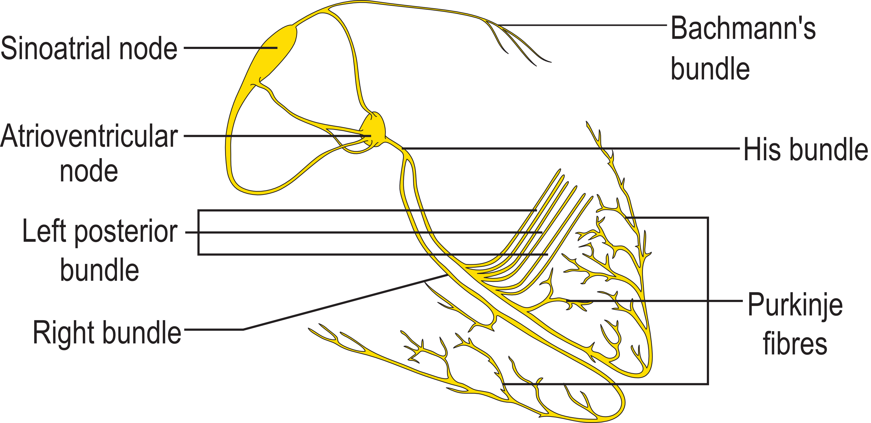
Normal Anatomy
- The conduction system of the heart consists of specialized cells designed to conduct electrical impulse faster than the surrounding myocardial cells.
- Anatomically, the AV node is divided into three regions as follows:
- Transitional cell zone: This is the region where the internodal atrial pathways merge with the compact AV node.
- Compact AV node: This region is located at the apex of the triangle of Koch, which is formed by the ostium of coronary sinus, tricuspid annulus and the tendon of Todaro.
- Penetrating portion of the AV bundle: This region enters the tendon of Todaro and runs within the fibrous body of the membranous interventricular septum and eventually divides at the crest of the muscular interventricular septum into right and left branches.
- The left bundle branch penetrates the membranous portion of the interventricular septum and divides into several smaller branches. Parts of the left bundle branch include a pre-divisional segment, anterior fascicle/hemibundle and posterior fascicle/hemibundle. Rarely a median fascicle is present in some hearts.
- The anterior fascicle supplies the anterior papillary muscle and the Purkinje network of the antero-lateral surface of the left ventricle.
- The posterior fascicle supplies the posterior papillary muscle and the Purkinje network of the postero-inferior surface of the left ventricle.
- Left bundle branch receives its blood supply from left anterior descending artery.

Normal Physiology
- The normal cardiac conduction proceeds in a way so as to allow time for the atrium to relax during atrial diastole.
- The electrical impulse generated in the SA node travels through the internodal pathways towards the AV node.
- The conduction through the AV node is slowed down as it travels through it. This decrease in velocity of conduction allows time for the atrium to contract ahead of the ventricle so that the blood from the atria can fill up the ventricles through the atrio-ventricular valves.
- As the impulse flows through the compact AV node, it rapidly conducts through the ventricular myocardial cells. Once the depolarization is complete, the ventricle relaxes during diastole in preparation for the next impulse.
Classification
Diagnosis
Electrocardiogram
A bundle branch block can be diagnosed when the duration of the QRS complex on the ECG exceeds 120 ms. A right bundle branch block typically causes prolongation of the last part of the QRS complex, and may shift the heart's electrical axis slightly to the right. The ECG will show a terminal R wave in lead V1 and a slurred S wave in lead I. Left bundle branch block widens the entire QRS, and in most cases shifts the heart's electrical axis to the left. The ECG will show a QS or rS complex in lead V1 and a monophasic R wave in lead I. Another normal finding with bundle branch block is appropriate T wave discordance. In other words, the T wave will be deflected opposite the terminal deflection of the QRS complex.
Treatment
Many people with bundle branch blocks may still be quite active, and may have nothing more remarkable than an abnormal appearance to their ECG. However, when bundle blocks are complex and diffuse in the bundle systems, or associated with additional and significant ventricular muscle damage, they may be a sign of serious underlying heart disease. In more severe cases, a pacemaker may be required to re-establish better heart muscle function.
Related Chapters
- Electrical conduction system of the heart
- Cardiac pacemaker
- Heart blocks
- First degree AV block
- Second degree AV block
- Third degree AV block
- Right bundle branch block
- Left bundle branch block
- Bifascicular block
- Trifascicular block