Accelerated idioventricular rhythm: Difference between revisions
No edit summary |
No edit summary |
||
| Line 17: | Line 17: | ||
'''Associate Editor-In-Chief:''' {{CZ}} | '''Associate Editor-In-Chief:''' {{CZ}} | ||
== | ==[[Accelerated idioventricular rhythm overview|Overview]]== | ||
The incidence of Accelerated Idioventricular Rhythms following acute MI is reported to be between 8 and 36%. This rhythm can also be seen in patients with primary myocardial disease, hypertensive, rheumatic, and congenital heart disease. Digoxin may cause of accelerated idioventricular rhythm. | The incidence of Accelerated Idioventricular Rhythms following acute MI is reported to be between 8 and 36%. This rhythm can also be seen in patients with primary myocardial disease, hypertensive, rheumatic, and congenital heart disease. Digoxin may cause of accelerated idioventricular rhythm. | ||
Revision as of 16:39, 22 August 2012
| Accelerated idioventricular rhythm | |
 | |
|---|---|
| 12 lead EKG shows a slow and wide complexes with intermittent narrow complex beats. The 5th and 10th beats are sinus node originated complexes (sinus rhythm) and close examination of these beats will give a clue to understand the cause of the wide complex rhythm. ST elevation indicates an acute myocardial infarction. The wide complex beats represent accelerated idioventricular rhythm which is usually seen following reperfusion in acute MI | |
| MeSH | D016170 |
|
Accelerated idioventricular rhythm Microchapters |
|
Differentiating Accelerated idioventricular rhythm from other Diseases |
|---|
|
Diagnosis |
|
Treatment |
|
Accelerated idioventricular rhythm On the Web |
|
American Roentgen Ray Society Images of Accelerated idioventricular rhythm |
|
Directions to Hospitals Treating Accelerated idioventricular rhythm |
|
Risk calculators and risk factors for Accelerated idioventricular rhythm |
| Cardiology Network |
 Discuss Accelerated idioventricular rhythm further in the WikiDoc Cardiology Network |
| Adult Congenital |
|---|
| Biomarkers |
| Cardiac Rehabilitation |
| Congestive Heart Failure |
| CT Angiography |
| Echocardiography |
| Electrophysiology |
| Cardiology General |
| Genetics |
| Health Economics |
| Hypertension |
| Interventional Cardiology |
| MRI |
| Nuclear Cardiology |
| Peripheral Arterial Disease |
| Prevention |
| Public Policy |
| Pulmonary Embolism |
| Stable Angina |
| Valvular Heart Disease |
| Vascular Medicine |
Editor-In-Chief: C. Michael Gibson, M.S., M.D. [1]
Associate Editor-In-Chief: Cafer Zorkun, M.D., Ph.D. [2]
Overview
The incidence of Accelerated Idioventricular Rhythms following acute MI is reported to be between 8 and 36%. This rhythm can also be seen in patients with primary myocardial disease, hypertensive, rheumatic, and congenital heart disease. Digoxin may cause of accelerated idioventricular rhythm.
EKG characteristics
- Regular rhythm at a rate of 40 to 100-120 BPM.
- QRS complexes are abnormal and wide.
- The ventricular complexes are usually but not necessarily dissociated from the P waves.
- Ventricular capture and fusion beats are common.
Differential Diagnosis
- Because of its slower rate it may resemble NSR. Look for numerous fusion beats. The term accelerated isorhythmic ventricular rhythm has been suggested.
- Must be distinguished from junctional tachycardia with preexisting IVCDs. But in these patients there are no fusion or capture beats.
Clinical Correlation
- Seen in both AMIs and IMIs.
- Commonly seen following reperfusion.
- Usually occurs during sinus bradycardia.
- May also be caused by digitalis.
AIVR appears similar to ventricular tachycardia but is benign and doesn't need any treatment.
References
- Hammill S. C. Electrocardiographic diagnoses: Criteria and definitions of abnormalities, Chapter 18, MAYO Clinic, Concise Textbook of Cardiology, 3rd edition, 2007 ISBN 0-8493-9057-5
- Engelen DJ, Gressin V, Krucoff MW, Theuns DA, Green C, Cheriex EC, Maison-Blanche P, Dassen WR, Wellens HJ, and Gorgels AP. Usefulness of frequent arrhythmias after epicardial recanalization in anterior wall acute myocardial infarction as a marker of cellular injury leading to poor recovery of left ventricular function. Am J Cardiol 2003 Nov 15; 92(10) 1143-9. PMID 14609586.
- Ilia R, Zahger D, Cafri C, Abu-Ful A, Weinstein JM, Yaroslavtsev S, Gilutz H, and Amit G. Predicting survival with reperfusion arrhythmias during primary percutaneous coronary intervention for acute myocardial infarction. Isr Med Assoc J 2007 Jan; 9(1) 21-3. PMID 17274350
- http://www.emedicine.com/med/topic12.htm
- Hampton, John (1997). The ECG Made Easy.
Additional resources
- ECGpedia: Course for interpretation of ECG
- The whole ECG - A basic ECG primer
- 12-lead ECG library
- Simulation tool to demonstrate and study the relation between the electric activity of the heart and the ECG
- ECG information from Children's Hospital Heart Center, Seattle
- National Heart, Lung, and Blood Institute, Diseases and Conditions Index
- A history of electrocardiography
- EKG Interpretations in infants and children
Examples
-
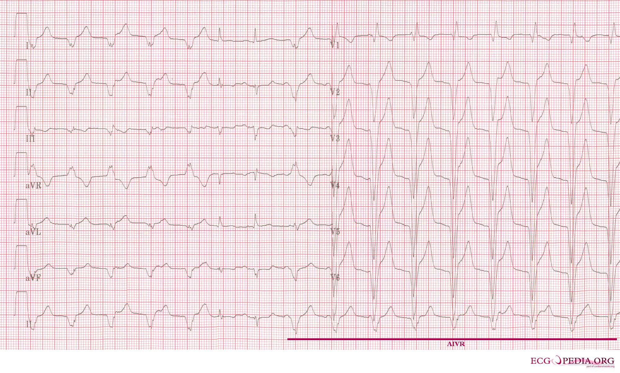
An example of accelerated idioventricular rhythm (AIVR)
-
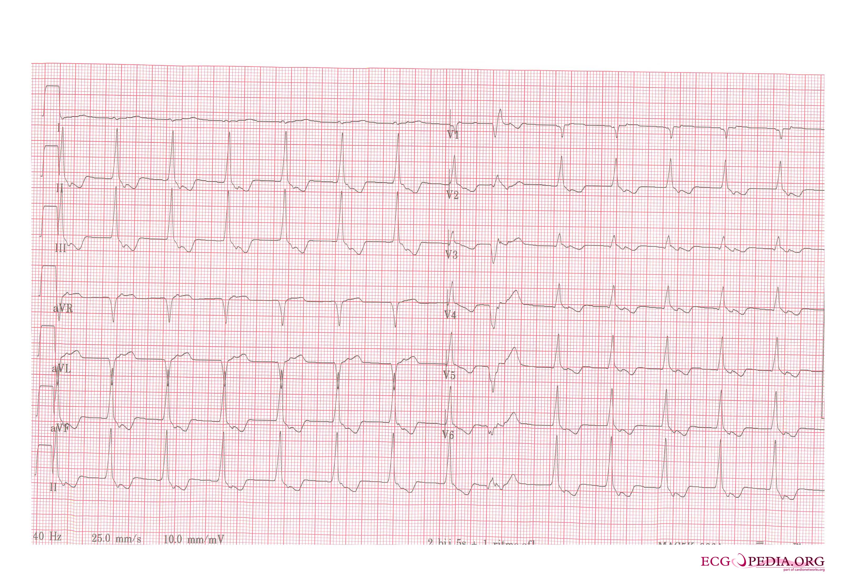
AIVR. Inverted P waves are sign of retrograde atrial activation.

