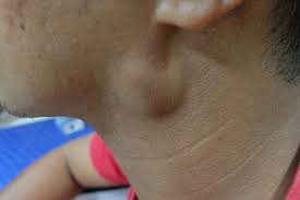
Lymphadenopathy overview

|
Lymphadenopathy Microchapters |
|
Diagnosis |
|---|
|
Treatment |
|
Case Studies |
|
Lymphadenopathy overview On the Web |
|
American Roentgen Ray Society Images of Lymphadenopathy overview |
|
Risk calculators and risk factors for Lymphadenopathy overview |
Editor-In-Chief: C. Michael Gibson, M.S., M.D. [1]; Associate Editor(s)-in-Chief: Amandeep Singh M.D.[2],, Raviteja Guddeti, M.B.B.S. [3]
Overview
Lymphadenopathy (also known as "enlarged lymph nodes") refers to lymph nodes which are abnormal in size, number, or consistency. Common causes of lymphadenopathy are infection, autoimmune disease, or malignancy. Lymphadenopathy may be classified according to distribution into 2 groups: generalized lymphadenopathy and localized lymphadenopathy. The pathogenesis of lymphadenopathy is characterized by the inflammation of lymph nodes. This process is primarily due to an elevated rate of trafficking of lymphocytes into the node from the blood, exceeding the rate of outflow from the node. Lymph nodes may also be enlarged secondarily as a result of the activation and proliferation of antigen-specific T and B cells (clonal expansion). Lymphadenopathy is very common, the estimated incidence of lymphadenopathy among children in the United States ranges from 35%- 45%. Patients of all age groups may develop lymphadenopathy. Lymphadenopathy is more commonly observed among children. Common complications of lymphadenopathy, may include: abscess formation, superior vena cava syndrome, and intestinal obstruction. Diagnostic criteria for malignant lymphadenopathy, may include: node > 2 cm, node that is draining, hard, or fixed to underlying tissue, atypical location (e.g. supraclavicular node), associated risk factors (e.g. HIV or TB), fever and/or weight loss, and splenomegaly. On the other hand, diagnostic criteria for benign lymphadenopathy, may include: node < 1 cm, node that is mobile, soft-or tender, and is not fixed to underlying tissue, typical location (e.g. supraclavicular node), no associated risk factors, and palpable and painful enlargement. Laboratory findings consistent with the diagnosis of lymphadenopathy may include elevated lactate dehydrogenase (LDH), mild neutropenia, and leukocytosis. There is no treatment for lymphadenopathy; the mainstay of therapy is treating the underlying condition.
Historical Perspective
Classification
Lymphadenopathy may be classified according to distribution into 2 groups localized lymphadenopathy and generalized lymphadenopathy. Lymphadenopathy may be classified as follows:
- By location:
- Dermatopathic lymphadenopathy: lymphadenopathy associated with skin disease.
- By malignancy: Benign lymphadenopathy is distinguished from malignant types which mainly refer to lymphomas or lymph node metastasis.
- By extent:
- Localized lymphadenopathy: due to localized spot of infection
- Generalized lymphadenopathy: due to systemic infection of the body. In some cases, it may persist for prolonged periods possibly without an apparent cause
- By size, where lymphadenopathy in adults is often defined as a short axis of one or more lymph nodes is greater than 10mm.
Pathophysiology
Lymph nodes are part of the immune system. As such, they are most readily palpable when fighting infections. Infections can either originate from the organs that they drain or primarily within the lymph node itself, referred to as lymphadenitis.*The pathogenesis of lymphadenopathy is characterized by the inflammation of lymph nodes. This process is primarily due to an elevated rate of trafficking of lymphocytes into the node from the blood, exceeding the rate of outflow from the node.[1]
- The immune response between the antigen and lymphocyte that leads to cellular proliferation and enlargement of the lymph nodes.
- Lymph nodes may also be enlarged secondarily as a result of the activation and proliferation of antigen-specific T and B cells (clonal expansion).
- On gross pathology, characteristic findings of lymphadenopathy, include:
- Enlarged lymph node
- Soft greasy yellow areas within the capsule
Lymph nodes are a part of the reticuloendothelial (RES) system, which includes lymphatic vessels, the lymphatic fluid found in interstitial fluid, monocytes of the blood, macrophages of the connective tissue, bone marrow, thymus, spleen, bone, and mucosa-associated lymphoid tissue (MALT) of visceral organs [1]
Lymphatic fluid moves throughout the lymphatic system and enters lymph nodes for filtration of foreign antigen. Foreign antigens are presented to the lymphoid cells, which lead to cellular proliferation and enlargement. Under microscopy, cellular proliferation in lymphoid follicles may be identified as several mitotic figures.[2] Increased activity leads to stretching of the lymphatic capsule and this may cause localized tenderness.
The development of B-cells originates from pluripotent stem cells from the bone marrow. B cells that successfully build their immunoglobulin heavy chains migrate to the germinal centers to allow for antibody diversification by somatic hypermutation.[3] The current school of thought is that B-cell lymphomas occur as a result of alternations in chromosomal translocations and somatic hypermutation.
T-cell development also begins from pluripotent stem cells, which mature within the thymic cortex. [4] While they are in the thymic cortex, specific rearrangements occur at the T-cell receptor. It is understood that chromosomal translocations at the level of T-cell receptors lead to T-cell lymphomagenesis.
Lymph nodes follicle necrosis may occur due to inflammatory, infectious, or malignant conditions. The neutrophil-rich infiltrates suggests bacterial infection, while lymphocyte-rich predominance may suggest viral infection. However, clinicians must remember that etiologies may vary; lymphomas, leukemias, tuberculosis, or even systemic lupus erythematosus (SLE) may be more appropriate diagnoses in the appropriate clinical context [5]
Causes
The most common causes of lymphadenopathy include infections, cancers, and connective tissue disorders. Lymph node enlargement can be of viral, bacterial, malignant, protozoan origin and can even be caused by live vaccines [6] Examples of infections that can cause lymph node enlargement include:
- Viral infections such as Epstein-Barr Virus and cytomegalovirus which cause infectious mononucleosis, [7] and CMV mononucleosis respectively.[8]
- Yersinia pestis, which causes the bubonic plague, causes lymph node swelling so large that it can be seen under the skin. These lymph nodes are called buboes and may become necrotic. [11]
- Other bacterial infections such as cat-scratch disease, [12] cutaneous anthrax, [13] and tuberculous lymphadenitis [14]
- Protozoal infections including African sleeping sickness, [15] Chagas' Disease, [16] and toxoplasmosis. [17]
Examples of malignancies that cause lymphadenopathy are:
- Primary: Hodgkin lymphoma [18] and non-Hodgkin lymphoma give lymphadenopathy in all or a few lymph nodes.[19]
- Secondary: metastasis, Virchow's Node, neuroblastoma, [20] and chronic lymphocytic leukemia.[21]
Autoimmune causes include: systemic lupus erythematosus [22] and rheumatoid arthritis may have a generalized lymphadenopathy.[19]
Benign lymphadenopathy
Examples include:
- Reactive Follicular hyperplasia [23]
- Atypical Follicular Hyperplasia [23]
- IgG4-related sclerosing disease-associated lymphadenopathy [23]
- Paracortical hyperplasia/Interfollicular hyperplasia: It is seen in viral infections, skin diseases, and nonspecific reactions. [23]
- Sinus histiocytosis: It is seen in lymph nodes draining limbs, inflammatory lesions, and malignancies. [23]
- Benign lymphadenopathy with extensive necrosis [23]
Axillary lymphadenopathy can be defined as solid nodes measuring more than 15 mm without fatty hilum.[24] Axillary lymph nodes may be normal up to 30 mm if consisting largely of fat.[24]
In children, a short axis of 8 mm can be used.[25] However, inguinal lymph nodes of up to 15 mm and cervical lymph nodes of up to 20 mm are generally normal in children up to age 8–12.[26]
Lymphadenopathy of more than 1.5 cm - 2 cm increases the risk of cancer or granulomatous disease as the cause rather than only inflammation or infection. Still, an increasing size and persistence over time are more indicative of cancer.[27]
Differentiating Lymphadenopathy from Other Diseases
Lymphadenopathy must be differentiated from syphilis, which may present as fever, myalgias, weight loss, and lymph node enlargement. After a thorough history and physical examination, lymphadenopathy can be initially categorized as:
Diagnostic: wherein the practitioner has a proximal cause for the lymph nodes and can go on to treat them. Examples would be Strep pharyngitis or localized cellulitis. The lymphadenopathy pattern history and physical examination can be suggestive an example would be mononucleosis wearing the practitioner has a strong clinic index of suspicion can perform a confirmatory test which if positive he can go on and treat the patient.
Unexplained lymphadenopathy. Unexplained lymphadenopathy can be generalized into localized or generalized lymphadenopathy. Unexplained localized lymphadenopathy is further divided into patterns at no risk for malignancy or severe illness in which case the patient can be observed for 3 to 4 weeks and if a response or improvement can be followed. The other alternative is if the patient is found to have a risk for malignancy or serious illness biopsy is indicated
Unexplained generalized lymphadenopathy can be approached after a review of epidemiological clues and medications with initial testing with a CBC with manual differential and mononucleosis serology if either is positive and diagnostic proceed to treatment. If both are negative, the second workup approach would be a PPD, and RPR, a chest x-ray, and ANA, hepatitis BS antigen serology and HIV. Additional testing modalities and lab tests may be indicated depending on clinical cues. If the results of this testing are conclusive, the practitioner can proceed on to diagnosis and treatment of the illness. If the results of the testing are still not clear, proceed to biopsy of the most abnormal of the nodes. The most functional way to investigate the differential diagnosis of lymphadenopathy is to characterize it by node pattern and location, obtained pertinent history including careful evaluation of epidemiology, and place the patient in the appropriate arm of the algorithm to evaluate lymphadenopathy.
Epidemiology and Demographics
The estimated incidence of lymphadenopathy in children in the United States ranges from 35%- 45%. It is more common in the pediatric population. Race and gender have no predilection in lymphadenopathy incidence.
Risk Factors
Common risk factors in the development of lymphadenopathy may be occupational, environmental, genetic, and viral.
Screening
There is insufficient evidence to recommend routine screening for lymphadenopathy
Natural History, Complications, and Prognosis
The natural course of lymphadenopathy depends on the underlying cause. Lymphadenopathy due to infectious causes subsides once the infection is controlled. Common complications of lymphadenopathy depend on the site of involvement, e.g. mediastinal lymphadenopathy include compression symptoms likeTracheal and bronchial obstruction and Dysphagia in Superior vena cava syndrome. Prognosis is generally excellent for infectious causes. Prompt treatment with antibiotics usually leads to a complete recovery. However, it may take weeks, or even months, for swelling to disappear. The amount of time to recovery depends on the cause. Prognosis is poor for malignant tumors.
Diagnosis
Diagnostic Criteria
Malignant Lymphadenopathy
- Node > 2 cm
- Node that is draining, hard, or fixed to underlying tissue
- Atypical location (e.g. supraclavicular node)
- Risk factors (e.g. HIV or TB)
- Fever and/or weight loss
- Splenomegaly
Benign Lymphadenopathy
- Node < 1 cm
- Node that is mobile, soft-or tender, and is not fixed to underlying tissue
- Common location (e.g. supraclavicular node)
- No associated risk factors
- Palpable and painful enlargement
History and Symptoms
The hallmark of lymphadenopathy is swollen lymph node. A positive history of a lump in the neck, red, tender skin over lymph node, and swollen, tender, or hard lymph nodes is suggestive of lymphadenopathy. The most common symptoms of lymphadenopathy include a lump in neck or affected part and constitutional symptoms like fatigue, fever, malaise, flu- like illness, nausea and vomiting, night sweats, weight loss, and cachexia.
Physical Examination
Common physical examination findings of lymphadenopathy include fever and tachycardia in infectious causes. There is an enlargement of different groups of lymph node chains depending upon the site of involvement and underlying causes.
Laboratory Findings
Electrocardiogram
X-ray
Echocardiography and Ultrasound
CT scan
MRI
Other Imaging Findings
Other Diagnostic Studies
Treatment
Treatment of lymphadenopathy is based on the etiology. Generally, treatment of lymphadenopathy is as follows:
- Infectious causes of lymphadenopathy can be treated with antibiotic therapy, antiviral therapy, or antifungal therapy.
- Immune therapy, systemic glucocorticoids can be used for autoimmune causes of lymphadenopathy
- For malignancies, any combination of surgery, chemotherapy, and radiation therapy can be used.
- If medication is the suspected cause, discontinue the medication if possible.
Medical Therapy
The medical therapy depends upon the underlying cause. Appropriate antibiotics are given for infective causes. Glucocorticoids for autoimmune conditions like sarcoidosis, and chemotherapy and radiation for malignant causes.
Surgery
Surgery is not the first-line treatment option for patients with lymphadenopathy. Surgery is usually reserved for patients with either malignancy and an indication of biopsy. It involves the removal or aspiration of lymph nodes. They are dissected when the cancer is in an advanced stage.
Primary Prevention
Good general health and hygiene are helpful in the prevention of any infection.
Secondary Prevention
Effective measures for the secondary prevention of lymphadenopathy include sentinel lymph node biopsy and early treatment if metastasis is detected.
References
- ↑ 1.0 1.1 Mohseni S, Shojaiefard A, Khorgami Z, Alinejad S, Ghorbani A, Ghafouri A (2014). "Peripheral lymphadenopathy: approach and diagnostic tools". Iran J Med Sci. 39 (2 Suppl): 158–70. PMC 3993046. PMID 24753638.
- ↑ Gowing NF (1974). "Tumours of the lymphoreticular system: nomenclature, histogenesis, and behaviour". J Clin Pathol Suppl (R Coll Pathol). 7: 103–7. PMC 1347234. PMID 4598345.
- ↑ Mesin L, Ersching J, Victora GD (2016). "Germinal Center [[B Cell]] Dynamics". Immunity. 45 (3): 471–482. doi:10.1016/j.immuni.2016.09.001. PMC 5123673. PMID 27653600. URL–wikilink conflict (help)
- ↑ Kumar BV, Connors TJ, Farber DL (2018) Human T Cell Development, Localization, and Function throughout Life. Immunity 48 (2):202-213. DOI:10.1016/j.immuni.2018.01.007 PMID: 29466753
- ↑ Strickler JG, Warnke RA, Weiss LM (1987). "Necrosis in lymph nodes". Pathol Annu. 22 Pt 2: 253–82. PMID 3317224.
- ↑ (2015) Reorganized text. JAMA Otolaryngol Head Neck Surg 141 (5):428. DOI:10.1001/jamaoto.2015.0540 PMID: 25996397
- ↑ Weiss LM, O'Malley D (2013). "Benign lymphadenopathies". Mod Pathol. 26 Suppl 1: S88–96. doi:10.1038/modpathol.2012.176. PMID 23281438.
- ↑ Sinha AK, Lovett M, Pillay G (1970). "Cytomegalovirus infection with Lymphadenopathy". Br Med J. 3 (5715): 163. doi:10.1136/bmj.3.5715.163. PMC 1702272. PMID 4317237.
- ↑ O'Leary J, Kennedy M, Howells D, Silva I, Uhlmann V, Luttich K; et al. (2000). "Cellular localisation of HHV-8 in Castleman's disease: is there a link with lymph node vascularity?". Mol Pathol. 53 (2): 69–76. doi:10.1136/mp.53.2.69. PMC 1186908. PMID 10889905.
- ↑ Oksenhendler E, Duarte M, Soulier J, Cacoub P, Welker Y, Cadranel J; et al. (1996). "Multicentric Castleman's disease in HIV infection: a clinical and pathological study of 20 patients". AIDS. 10 (1): 61–7. PMID 8924253.
- ↑ Butler T (2009). "Plague into the 21st century". Clin Infect Dis. 49 (5): 736–42. doi:10.1086/604718. PMID 19606935.
- ↑ Klotz SA, Ianas V, Elliott SP (2011). "Cat-scratch Disease". Am Fam Physician. 83 (2): 152–5. PMID 21243990.
- ↑ Sweeney DA, Hicks CW, Cui X, Li Y, Eichacker PQ (2011). "Anthrax infection". Am J Respir Crit Care Med. 184 (12): 1333–41. doi:10.1164/rccm.201102-0209CI. PMC 3361358. PMID 21852539.
- ↑ Fontanilla JM, Barnes A, von Reyn CF (2011). "Current diagnosis and management of peripheral tuberculous lymphadenitis". Clin Infect Dis. 53 (6): 555–62. doi:10.1093/cid/cir454. PMID 21865192.
- ↑ Kennedy PG (2013) Clinical features, diagnosis, and treatment of human African trypanosomiasis (sleeping sickness). Lancet Neurol 12 (2):186-94. DOI:10.1016/S1474-4422(12)70296-X PMID: 23260189
- ↑ Salazar Schettino PM, Bucio Torres M, Cabrera Bravo M, Ruiz Hernández AL (2011). "[Chagas disease in Mexico. Report of two acute cases]". Gac Med Mex. 147 (1): 63–9. PMID 21412398.
- ↑ Montoya JG, Liesenfeld O (2004). "Toxoplasmosis". Lancet. 363 (9425): 1965–76. doi:10.1016/S0140-6736(04)16412-X. PMID 15194258.
- ↑ Glass, C (September 2008). "Role of the Primary Care Physician in Hodgkin Lymphoma". American Family Physician. 78 (5): 615–622. PMID 18788239.
- ↑ 19.0 19.1 Status and anamnesis, Anders Albinsson. Page 12
- ↑ Colon, NC; Chung, DH (2011). "Neuroblastoma". Advances in Pediatrics. 58 (1): 297–311. doi:10.1016/j.yapd.2011.03.011. PMC 3668791. PMID 21736987.
- ↑ Sagatys, EM; Zhang, L (January 2011). "Clinical and laboratory prognostic indicators in chronic lymphocytic leukemia". Cancer Control. 19 (1): 18–25. doi:10.1177/107327481201900103. PMID 22143059.
- ↑ Melikoglu, MA; Melikoglu, M (October–December 2008). "The clinical importance of lymphadenopathy in systemic lupus erythematosus" (PDF). Acta Reumatologia Portuguesa. 33 (4): 402–406. PMID 19107085.
- ↑ 23.0 23.1 23.2 23.3 23.4 23.5 Weiss, LM; O'Malley, D (2013). "Benign lymphadenopathies". Modern Pathology. 26 (Supplement 1): S88–S96. doi:10.1038/modpathol.2012.176. PMID 23281438.
- ↑ 24.0 24.1 Page 559 in: Wolfgang Dähnert (2011). Radiology Review Manual. Lippincott Williams & Wilkins. ISBN 9781609139438.
- ↑ Page 942 in: Richard M. Gore, Marc S. Levine (2010). High Yield Imaging Gastrointestinal HIGH YIELD in Radiology. Elsevier Health Sciences. ISBN 9781455711444.
- ↑ Laurence Knott. "Generalised Lymphadenopathy". Patient UK. Retrieved 2017-03-04. Last checked: 24 March 2014
- ↑ Bazemore AW, Smucker DR (December 2002). "Lymphadenopathy and malignancy". American Family Physician. 66 (11): 2103–10. PMID 12484692.