Esophageal varices: Difference between revisions
m (Bot: Automated text replacement (-{{SIB}} + & -{{EJ}} + & -{{EH}} + & -{{Editor Join}} + & -{{Editor Help}} +)) |
No edit summary |
||
| Line 1: | Line 1: | ||
__NOTOC__ | |||
{{Infobox_Disease | | {{Infobox_Disease | | ||
Name = Esophageal varices | | Name = Esophageal varices | | ||
| Line 14: | Line 10: | ||
OMIM = | | OMIM = | | ||
MedlinePlus = 000268 | | MedlinePlus = 000268 | | ||
eMedicineSubj = | eMedicineSubj = | | ||
eMedicineTopic = | eMedicineTopic = | | ||
eMedicine_mult = | eMedicine_mult = | | ||
MeshID = D004932 | | MeshID = D004932 | | ||
}} | }} | ||
{{Search infobox}} | {{Search infobox}} | ||
{{CMG}} | |||
==Overview== | |||
In [[medicine]] ([[gastroenterology]]), '''esophageal varices''' are extremely [[dilation|dilated]] sub-mucosal [[vein]]s in the [[esophagus]]. They are most often a consequence of [[portal hypertension]], such as may be seen with [[cirrhosis]]; patients with esophageal varices have a strong tendency to develop [[bleeding]]. | In [[medicine]] ([[gastroenterology]]), '''esophageal varices''' are extremely [[dilation|dilated]] sub-mucosal [[vein]]s in the [[esophagus]]. They are most often a consequence of [[portal hypertension]], such as may be seen with [[cirrhosis]]; patients with esophageal varices have a strong tendency to develop [[bleeding]]. | ||
| Line 34: | Line 33: | ||
Varices can also form in other areas of the body, including the [[stomach]] ('''[[gastric varices]]'''), [[duodenum]] ('''[[intestinal varices|duodenal varices]]'''), and [[rectum]] ('''[[rectal varices]]'''). Treatment of these types of varices may differ. | Varices can also form in other areas of the body, including the [[stomach]] ('''[[gastric varices]]'''), [[duodenum]] ('''[[intestinal varices|duodenal varices]]'''), and [[rectum]] ('''[[rectal varices]]'''). Treatment of these types of varices may differ. | ||
==Treatment | ==Treatment== | ||
In emergency situations, the care is directed at stopping blood loss, maintaining plasma volume, correcting disorders in coagulation induced by cirrhosis, and appropriate use of [[antibiotics]] (as infection is either concomitant, or a precipitant). | In emergency situations, the care is directed at stopping blood loss, maintaining plasma volume, correcting disorders in coagulation induced by cirrhosis, and appropriate use of [[antibiotics]] (as infection is either concomitant, or a precipitant). | ||
Revision as of 14:50, 6 September 2012
| Esophageal varices | |
 | |
|---|---|
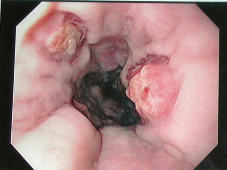
| Gastroscopy image of esophageal varices with prominent red wale spots | |
| ICD-10 | I85 |
| ICD-9 | 456.0-456.2 |
| DiseasesDB | 9177 |
| MedlinePlus | 000268 |
| MeSH | D004932 |
Template:Search infobox Editor-In-Chief: C. Michael Gibson, M.S., M.D. [1]
Overview
In medicine (gastroenterology), esophageal varices are extremely dilated sub-mucosal veins in the esophagus. They are most often a consequence of portal hypertension, such as may be seen with cirrhosis; patients with esophageal varices have a strong tendency to develop bleeding.
Esophageal varices are diagnosed with endoscopy.[1]
Pathogenesis
The majority of blood from the esophagus is drained away via the esophageal veins, which drain deoxygenated blood from the esophagus to the azygos vein which in turn, directly drains into the superior vena cava. These veins have no part in the development of esophageal varices. The remaining blood from the esophagus is drained away via the superficial veins lining the esophagus interior, which drain into the coronary vein (left gastric vein) which in turn, drains directly into the portal vein. These superficial veins lining the esophagus interior (normally only approximately 1mm in diameter) become distended up to 1-2 cm in diameter in association with portal hypertension.
Normal portal pressure is approximately 9 mmHg compared to an inferior vena cava pressure of 2-6 mmHg. This creates a normal pressure gradient of 3-7 mmHg. If the portal pressure rises above 12mmHg, this gradient rises to 7-10 mmHg.[2] A gradient greater than 10 mmHg is considered portal hypertension. At gradients greater than 10 mmHg, blood flow though the hepatic portal system is redirected from the liver into areas with lower venous pressures. This means that collateral circulation develops in the lower esophagus, abdominal wall, stomach and rectum. The small blood vessels in these areas become distended, becoming more thin-walled, and appear as varicosities. In addition, these vessels are poorly supported by other structures, as they are not designed for high pressures.
In situations where portal pressures increase, such as with cirrhosis, there is dilation of veins in the anastomosis, leading to esophageal varices.
Varices can also form in other areas of the body, including the stomach (gastric varices), duodenum (duodenal varices), and rectum (rectal varices). Treatment of these types of varices may differ.
Treatment
In emergency situations, the care is directed at stopping blood loss, maintaining plasma volume, correcting disorders in coagulation induced by cirrhosis, and appropriate use of antibiotics (as infection is either concomitant, or a precipitant).
Therapeutic endoscopy is considered the mainstay of urgent treatment. Two main therapeutic approaches exist:
- Variceal ligation, or banding
- sclerotherapy
In cases of refractory bleeding, balloon tamponade may be necessary, usually as a bridge to further endoscopy, a transjugular intrahepatic portosystemic shunt (TIPS), or a distal splenorenal shunt procedure or a liver transplantation.
Nutritional supplementation is not necessary if the patient is not eating for four days or less.[3]
-
Esophageal varices seven days post banding, showing ulceration at the site of banding.

Prevention
Ideally, patients with known varices should receive treatment to reduce their risk of bleeding.[4] The non-selective β-blockers (e.g., propranolol, timolol or nadolol) and nitrates have been evaluated for secondary prophylaxis. The effectiveness of this treatment has been shown by a number of different studies.[5]
Unfortunately, non-selective β-blockers do not prevent the formation of esophageal varices.
See also
References
- ↑ Biecker E, Schepke M, Sauerbruch T (2005). "The role of endoscopy in portal hypertension". Dig Dis. 23 (1): 11–7. PMID 15920321.
- ↑ Arguedas M (2003). "The critically ill liver patient: the variceal bleeder". Semin Gastrointest Dis. 14 (1): 34–8. PMID 12610853.
- ↑ de Lédinghen V, Beau P, Mannant PR; et al. (1997). "Early feeding or enteral nutrition in patients with cirrhosis after bleeding from esophageal varices? A randomized controlled study". Dig. Dis. Sci. 42 (3): 536–41. PMID 9073135.
- ↑ Lebrec D, Poynard T, Hillon P, Benhamou J-P (1981). "Propranolol for prevention of recurrent gastrointestinal bleeding in patients with cirrhosis: a controlled study". N Engl J Med. 305: 1371&ndash, 1374. PMID 7029276.
- ↑ Talwalkar JA, Kamath PS (2004). "An evidence-based medicine approach to beta-blocker therapy in patients with cirrhosis". Am J Med. 116: 759&ndash, 766. PMID 15144913.
See also