Carotid artery stenosis: Difference between revisions

Jump to navigation Jump to search
No edit summary
 
 
(28 intermediate revisions by 4 users not shown)
Line 5: Line 5:
   Image          = Carotid-artery-stenosis-001.jpg|
   Image          = Carotid-artery-stenosis-001.jpg|
   Caption        = |
   Caption        = |
  DiseasesDB    = 31178 |
  ICD10          = {{ICD10|I|65|2|i|60}} |
  ICD9          = {{ICD9|433.1}} |
  ICDO          = |
  OMIM          = |
  MedlinePlus    = |
  eMedicineSubj  = |
  eMedicineTopic = |
  MeshID        = D016893 |
}}
}}
{{SI}}
 
{{WikiDoc Cardiology Network Infobox}}
{{Carotid artery stenosis}}
 
{{CMG}}; '''Associate Editor-In-Chief:''' {{CZ}}


==Overview==
==[[Carotid artery stenosis overview|Overview]]==


'''Carotid arterial stenosis''' is a narrowing of the lumen of the [[carotid artery]], usually by [[atheroma]] (a fatty lump or plaque  causing[[ atherosclerosis]]). Atheroma's may cause [[transient ischemic attack]]s (TIAs) and [[cerebrovascular accident]]s (CVAs) as it obstructs the bloodstream to the [[brain]].  It also has the potential to generate [[embolus|emboli]] (blood clots) that obstruct the cerebral arteries.
==[[Carotid artery stenosis historical perspective|Historical Perspective]]==


The narrowing can either be [[asymptomatic]] (causing no medical problems) or presents with symptoms such as TIAs and CVAs.
==[[Carotid artery stenosis classification|Classification]]==


==Diagnosis==
==[[Carotid artery stenosis pathophysiology|Pathophysiology]]==
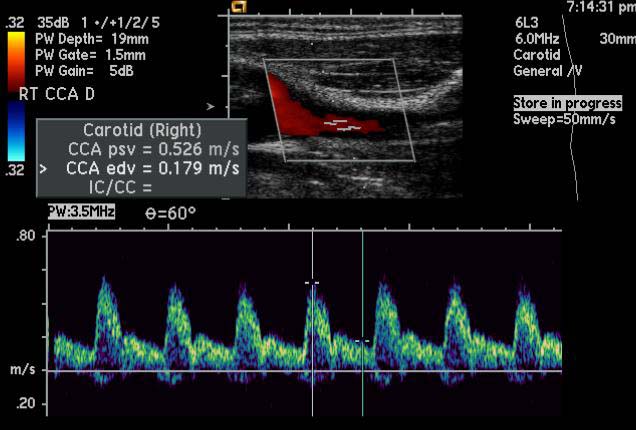
Carotid stenosis is usually diagnosed by [[ultrasound]] scan of the neck arteries.  This is the first imaging option and usually used for follow up and observation as it involves no radiation and no contrast agents that may cause [[allergic]] reactions.


If there is doubt whether the narrowing is still patent (open to blood flow) and the patient is to be assessed for treatment, the next imaging option would either be [[computed tomography]] [[angiogram]] (CTA) or a [[magnetic resonance imaging]] angiogram (MRA).
==[[Carotid artery stenosis causes|Causes]]==


CTA is usually used as it's more widely available and faster (takes only several minutes).  However, it does involve significant [[X-ray]] radiation and the use of [[contrast agent]]s which is usually iodinated, which can cause allergic reactions, even [[anaphylaxis]] and can also cause [[renal failure]].
==[[Carotid artery stenosis differential diagnosis|Differentiating Carotid artery stenosis from other Diseases]]==


MRA takes much longer to complete, more costly and not yet as widely available.
==[[Carotid artery stenosis epidemiology and demographics|Epidemiology and Demographics]]==


==Ultrasound Criteria for Carotid Artery Stenosis==
==[[Carotid artery stenosis risk factors|Risk Factors]]==
*Normal:
**ICA PSV is less than 125 cm/sec and no plaque or intimal thickening is visible sonographically.
**Additional criteria include ICA/CCA PSV ratio < 2.0 and ICA EDV < 40 cm/sec.
*<50% ICA stenosis:
**ICA PSV is less than 125 cm/sec and plaque or intimal thickening is visible sonographically.
**Additional criteria include ICA/CCA PSV ratio < 2.0 and ICA EDV < 40 cm/sec.
*50%–69% ICA stenosis:
**ICA PSV is 125–230 cm/sec and plaque is visible sonographically.
**Additional criteria include ICA/CCA PSV ratio of 2.0–4.0 and ICA EDV of 40–100 cm/sec.
*>=70% ICA stenosis but less than near occlusion
**ICA PSV is greater than 230 cm/sec and visible plaque and luminal narrowing are seen at gray-scale and color Doppler US (The higher the Doppler parameter lies above the threshold of 230 cm/sec, the greater the likelihood of severe disease).
**Additional criteria include ICA/CCA PSV ratio > 4 and ICA EDV > 100 cm/sec.
*Vear occlusion of the ICA:
**Velocity parameters may not apply, since velocities may be high, low, or undetectable.
**Diagnosis is established primarily by demonstrating a markedly narrowed lumen at color or power Doppler US (35).
*Total occlusion of the ICA
**No detectable patent lumen at gray-scale US and no flow with spectral, power, and color Doppler US. <ref>Edward G. Grant, Carol B. Benson, Gregory L. Moneta, Andrei V. Alexandrov, J. Dennis Baker, Edward I. Bluth, Barbara A. Carroll, Michael Eliasziw, John Gocke, Barbara S. Hertzberg, Sandra Katanick, Laurence Needleman, John Pellerito, Joseph F. Polak, Kenneth S. Rholl, Douglas L. Wooster, and Eugene Zierler. [http://radiology.rsnajnls.org/cgi/content/short/2292030516v1 Carotid Artery Stenosis: Gray-Scale and Doppler US Diagnosis—Society of Radiologists in Ultrasound Consensus Conference.] Radiology published online September 18, 2003.</ref>


==Treatment==
==[[Carotid artery stenosis screening|Screening]]==
Options for treatment include:
*Conservative: [[Antiplatelet drug]]
*[[Carotid endarterectomy]] (surgical removal of the atheroma)
*[[Carotid stenting]]


==Examples==
==[[Carotid artery stenosis natural history, complications and prognosis|Natural History, Complications and Prognosis]]==


<div align="left">
==Diagnosis==
<gallery heights="175" widths="175">
[[Carotid artery stenosis diagnostic testing guidelines|Diagnostic Testing Guidelines]] | [[Carotid artery stenosis history and symptoms|History and Symptoms]] | [[Carotid artery stenosis physical examination|Physical Examination]] | [[Carotid artery stenosis laboratory findings|Laboratory Findings]] | [[Carotid artery stenosis electrocardiogram|Electrocardiogram]] | [[Carotid artery stenosis CT|CT]] | [[Carotid artery stenosis MRI|MRI]] | [[Carotid artery stenosis MRA|MRA]] | [[Carotid artery stenosis echocardiography or ultrasound|Echocardiography or Ultrasound]] | [[Carotid artery stenosis other imaging findings|Other Imaging Findings]] | [[Carotid artery stenosis other diagnostic studies|Other Diagnostic Studies]]
Image:Carotid-artery-stenosis-001.jpg
Image:Carotid-artery-stenosis-002.jpg
Image:Carotid-artery-stenosis-003.jpg
</gallery>
</div>


==Treatment==


<youtube v=nkCmbY_oLAo/>
[[Carotid artery stenosis medical therapy|Medical Therapy]] | [[Carotid artery stenosis surgery|Surgery]] | [[Carotid artery stenosis primary prevention|Primary Prevention]] | [[Carotid artery stenosis secondary prevention|Secondary Prevention]] | [[Carotid artery stenosis cost-effectiveness of therapy|Cost-Effectiveness of Therapy]] | [[Carotid artery stenosis future or investigational therapies|Future or Investigational Therapies]]


==Case Studies==
[[Carotid artery stenosis case study one|Case #1]]


==See also==
==Related Chapters==
*[[Ocular ischemic syndrome]]
*[[External carotid artery]]
*[[Common carotid artery]]
*[[Internal carotid artery]]
*[[Carotid artery dissection]]
*[[Amaurosis fugax]]


==References==
{{Circulatory system pathology}}
{{Reflist|2}}


{{Circulatory system pathology}}
{{SIB}}


[[Category:Neurology]]
[[Category:Neurology]]
Line 87: Line 57:
[[Category:Emergency medicine]]
[[Category:Emergency medicine]]
[[Category:Overview complete]]
[[Category:Overview complete]]
 
[[Category:Disease]]
[[nl:Carotisstenose]]
[[nl:Carotisstenose]]


Latest revision as of 19:55, 30 January 2013

For patient information, click here

Carotid artery stenosis

Carotid artery stenosis Microchapters

Home

Patient Information

Overview

Historical Perspective

Classification

Pathophysiology

Causes

Differentiating Carotid artery stenosis from other Diseases

Epidemiology and Demographics

Risk Factors

Screening

Natural History, Complications and Prognosis

Diagnosis

Diagnostic Testing Guidelines

History and Symptoms

Physical Examination

Laboratory Findings

Electrocardiogram

CT

MRI

MRA

Echocardiography or Ultrasound

Other Imaging Findings

Other Diagnostic Studies

Treatment

Medical Therapy

Surgery

Primary Prevention

Secondary Prevention

Cost-Effectiveness of Therapy

Future or Investigational Therapies

ACC/AHA Guideline Recommendations

Diagnostic Testing Guidelines Recommendation

Primary Prevention and Screening Guidelines Recommendations

Secondary Prevention Guidelines Recommendations

Selection of Patients for Carotid Revascularization

Periprocedural Management of Patients Undergoing Carotid Endarterectomy

Management of Patients Undergoing Carotid Artery Stenting

Restenosis After Carotid Endarterectomy or Stenting

Vascular Imaging in Patients With Vertebral Artery Disease

Atherosclerotic Risk Factors in Patients With Vertebral Artery Disease

Occlusive Disease of the Subclavian and Brachiocephalic Arteries

Fibromuscular Dysplasia

Cervical Artery Dissection

Case Studies

Case #1

Carotid artery stenosis On the Web

Most recent articles

Most cited articles

Review articles

CME Programs

Powerpoint slides

Images

American Roentgen Ray Society Images of Carotid artery stenosis

All Images
X-rays
Echo & Ultrasound
CT Images
MRI

Ongoing Trials at Clinical Trials.gov

US National Guidelines Clearinghouse

NICE Guidance

FDA on Carotid artery stenosis

CDC on Carotid artery stenosis

Carotid artery stenosis in the news

Blogs on Carotid artery stenosis

Directions to Hospitals Treating Carotid artery stenosis

Risk calculators and risk factors for Carotid artery stenosis

Editor-In-Chief: C. Michael Gibson, M.S., M.D. [1]; Associate Editor-In-Chief: Cafer Zorkun, M.D., Ph.D. [2]

Overview

Historical Perspective

Classification

Pathophysiology

Causes

Differentiating Carotid artery stenosis from other Diseases

Epidemiology and Demographics

Risk Factors

Screening

Natural History, Complications and Prognosis

Diagnosis

Diagnostic Testing Guidelines | History and Symptoms | Physical Examination | Laboratory Findings | Electrocardiogram | CT | MRI | MRA | Echocardiography or Ultrasound | Other Imaging Findings | Other Diagnostic Studies

Treatment

Medical Therapy | Surgery | Primary Prevention | Secondary Prevention | Cost-Effectiveness of Therapy | Future or Investigational Therapies

Case Studies

Case #1

Related Chapters

nl:Carotisstenose


Template:WikiDoc Sources