Pulmonary vein stenosis
(Redirected from Pulmonary veno-occlusive disease)
| Pulmonary vein stenosis | |
 | |
|---|---|
| Pulmonary vein stenosis: Gross, natural color, close-up view, veins draining into atrium. Image courtesy of Professor Peter Anderson DVM PhD and published with permission © PEIR, University of Alabama at Birmingham, Department of Pathology |
Editor-In-Chief: C. Michael Gibson, M.S., M.D. [1]
Overview
Pulmonary vein stenosis is extremely rare, progressive and usually fatal, if left untreated.
Isolated stenosis of pulmonary veins, as they enter into the left atrium, is a rare congenital cardiac abnormality that has significant impact on clinical outcome.
Diagnosis is difficult to suspect, especially in the absence of other cardiac abnormalities.
Symptoms and clinical signs of pulmonary vein stenosis are very similar to those of chronic lung disease and are very difficult to differentiate.
Historical Perspective
- [Disease name] was first discovered by [scientist name], a [nationality + occupation], in [year] during/following [event].
- In [year], [gene] mutations were first identified in the pathogenesis of [disease name].
- In [year], the first [discovery] was developed by [scientist] to treat/diagnose [disease name].
Classification
- [Disease name] may be classified according to [classification method] into [number] subtypes/groups:
- [group1]
- [group2]
- [group3]
- Other variants of [disease name] include [disease subtype 1], [disease subtype 2], and [disease subtype 3].
Pathophysiology
Normal Pulmonary Vein (Lateral)
<googlevideo>-1618821798677201341&hl=en</googlevideo>
Normal Pulmonary Vein (PA)
<googlevideo>7010087554166470052&hl=en</googlevideo>
Left Upper Pulmonary Vein Stenosis Pulsed Wave Echo

Left Upper Pulmonary Vein Stenosis post Atrial Fibrillation Ablation
<googlevideo>-2781473498147468042&hl=en</googlevideo>
Pathological Findings
-
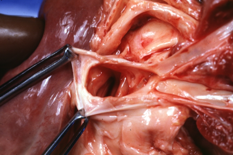 Pulmonary vein stenosis: Gross, natural color, close-up view of two veins entering atrium
 -
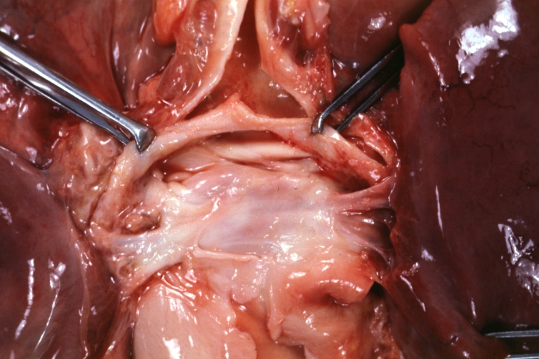
 Pulmonary vein stenosis: Gross, natural color, pulmonary outlet showing thickened pulmonary arteries


-
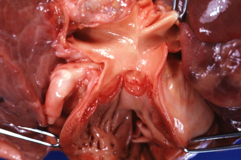
 Pulmonary vein stenosis: Gross, natural color, unopened veins and atrium dorsal view
 -
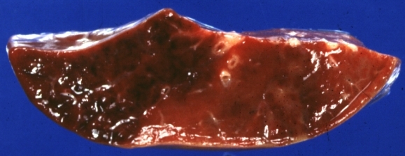 Pulmonary vein stenosis: Gross, natural color. cut surface. little to show except congestion and probable intrapulmonary hemorrhage


-
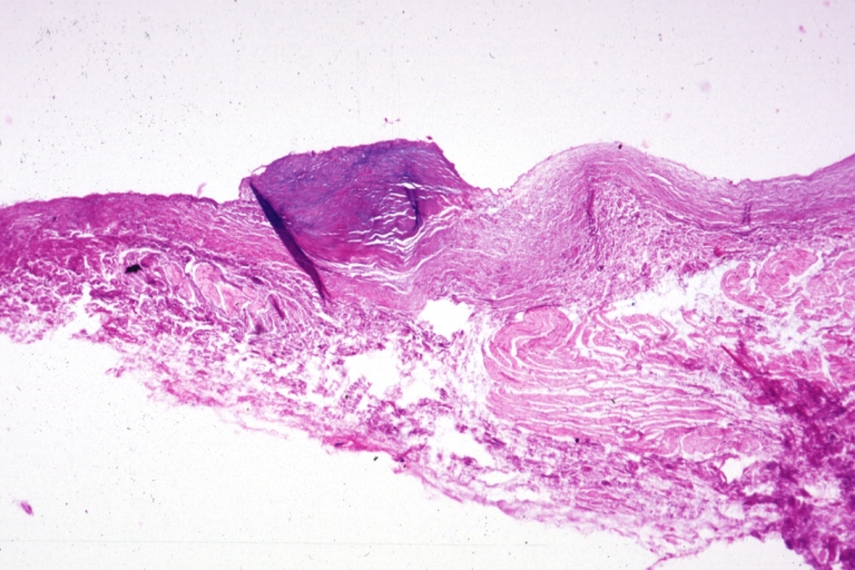 Pulmonary vein stenosis: Micro low mag, H&E, junction of pulmonary vein and atrium
 -
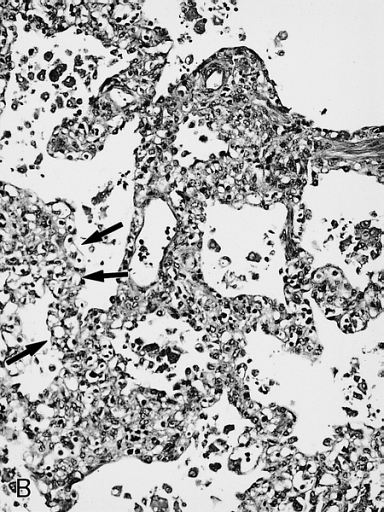
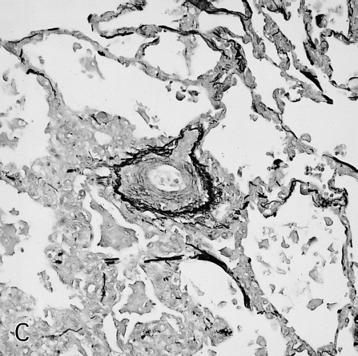
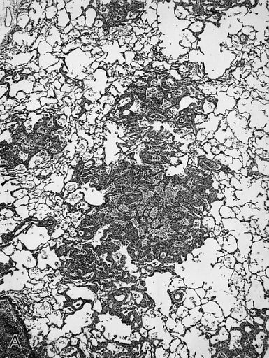 There is a patchy interstitial widening (A) due to proliferation of thin-walled capillaries in the alveolar walls (B, arrows). Secondary intimal thickening in a pulmonary vein is present (C, elastic tissue stain) indicative of secondary veno-occlusive disease and associated pulmonary hypertension.


-
 There is a patchy interstitial widening (A) due to proliferation of thin-walled capillaries in the alveolar walls (B, arrows). Secondary intimal thickening in a pulmonary vein is present (C, elastic tissue stain) indicative of secondary veno-occlusive disease and associated pulmonary hypertension.
 -
 There is a patchy interstitial widening (A) due to proliferation of thin-walled capillaries in the alveolar walls (B, arrows). Secondary intimal thickening in a pulmonary vein is present (C, elastic tissue stain) indicative of secondary veno-occlusive disease and associated pulmonary hypertension.


Causes
- [Disease name] may be caused by either [cause1], [cause2], or [cause3].
- [Disease name] is caused by a mutation in the [gene1], [gene2], or [gene3] gene[s].
- There are no established causes for [disease name].
Differentiating [disease name] from other Diseases
- [Disease name] must be differentiated from other diseases that cause [clinical feature 1], [clinical feature 2], and [clinical feature 3], such as:
- [Differential dx1]
- [Differential dx2]
- [Differential dx3]
Epidemiology and Demographics
- The prevalence of [disease name] is approximately [number or range] per 100,000 individuals worldwide.
- In [year], the incidence of [disease name] was estimated to be [number or range] cases per 100,000 individuals in [location].
Age
- Patients of all age groups may develop [disease name].
- [Disease name] is more commonly observed among patients aged [age range] years old.
- [Disease name] is more commonly observed among [elderly patients/young patients/children].
Gender
- [Disease name] affects men and women equally.
- [Gender 1] are more commonly affected with [disease name] than [gender 2].
- The [gender 1] to [Gender 2] ratio is approximately [number > 1] to 1.
Race
- There is no racial predilection for [disease name].
- [Disease name] usually affects individuals of the [race 1] race.
- [Race 2] individuals are less likely to develop [disease name].
Risk Factors
- Common risk factors in the development of [disease name] are [risk factor 1], [risk factor 2], [risk factor 3], and [risk factor 4].
Natural History, Complications and Prognosis
- The majority of patients with [disease name] remain asymptomatic for [duration/years].
- Early clinical features include [manifestation 1], [manifestation 2], and [manifestation 3].
- If left untreated, [#%] of patients with [disease name] may progress to develop [manifestation 1], [manifestation 2], and [manifestation 3].
- Common complications of [disease name] include [complication 1], [complication 2], and [complication 3].
- Prognosis is generally [excellent/good/poor], and the [1/5/10year mortality/survival rate] of patients with [disease name] is approximately [#%].
Diagnosis
Diagnostic Criteria
- The diagnosis of [disease name] is made when at least [number] of the following [number] diagnostic criteria are met:
- [criterion 1]
- [criterion 2]
- [criterion 3]
- [criterion 4]
Symptoms
- [Disease name] is usually asymptomatic.
- Symptoms of [disease name] may include the following:
- [symptom 1]
- [symptom 2]
- [symptom 3]
- [symptom 4]
- [symptom 5]
- [symptom 6]
Physical Examination
- Patients with [disease name] usually appear [general appearance].
- Physical examination may be remarkable for:
- [finding 1]
- [finding 2]
- [finding 3]
- [finding 4]
- [finding 5]
- [finding 6]
Laboratory Findings
- There are no specific laboratory findings associated with [disease name].
- A [positive/negative] [test name] is diagnostic of [disease name].
- An [elevated/reduced] concentration of [serum/blood/urinary/CSF/other] [lab test] is diagnostic of [disease name].
- Other laboratory findings consistent with the diagnosis of [disease name] include [abnormal test 1], [abnormal test 2], and [abnormal test 3].
Imaging Findings
- There are no [imaging study] findings associated with [disease name].
- [Imaging study 1] is the imaging modality of choice for [disease name].
- On [imaging study 1], [disease name] is characterized by [finding 1], [finding 2], and [finding 3].
- [Imaging study 2] may demonstrate [finding 1], [finding 2], and [finding 3].
Other Diagnostic Studies
- [Disease name] may also be diagnosed using [diagnostic study name].
- Findings on [diagnostic study name] include [finding 1], [finding 2], and [finding 3].
Treatment
Medical Therapy
- There is no treatment for [disease name]; the mainstay of therapy is supportive care.
- The mainstay of therapy for [disease name] is [medical therapy 1] and [medical therapy 2].
- [Medical therapy 1] acts by [mechanism of action 1].
- Response to [medical therapy 1] can be monitored with [test/physical finding/imaging] every [frequency/duration].
Surgery
- Surgery is the mainstay of therapy for [disease name].
- [Surgical procedure] in conjunction with [chemotherapy/radiation] is the most common approach to the treatment of [disease name].
- [Surgical procedure] can only be performed for patients with [disease stage] [disease name].
Prevention
- There are no primary preventive measures available for [disease name].
- Effective measures for the primary prevention of [disease name] include [measure1], [measure2], and [measure3].
- Once diagnosed and successfully treated, patients with [disease name] are followed-up every [duration]. Follow-up testing includes [test 1], [test 2], and [test 3].
References