Left coronary artery
|
Coronary Angiography | |
|
General Principles | |
|---|---|
|
Anatomy & Projection Angles | |
|
Normal Anatomy | |
|
Anatomic Variants | |
|
Projection Angles | |
|
Epicardial Flow & Myocardial Perfusion | |
|
Epicardial Flow | |
|
Myocardial Perfusion | |
|
Lesion Complexity | |
|
ACC/AHA Lesion-Specific Classification of the Primary Target Stenosis | |
|
Lesion Morphology | |
Editor-In-Chief: C. Michael Gibson, M.S., M.D. [1]; Associate Editor(s)-in-Chief: Rim Halaby, M.D. [2]; Hilda Mahmoudi M.D., M.P.H.[3]
Synonyms and keywords: LCA
Overview
The left coronary artery normally arises from the aortic sinus above the left cusp of the aortic valve. The origin of the left coronary artery is called the left main coronary artery, and it bifurcates into the left anterior descending and the left circumflex artery. This artery supplies the majority of the blood flow to the left ventricle, the pumping chamber of the heart. In fact, the LCA supplies the posterolateral side of the left ventricle as well as the anterior part of the left ventricle which includes the anterolateral myocardium, the apex, the anterior interventricular septum and the anterolateral papillary muscle.
Branches
The left coronary artery typically courses for 1 to 25 mm as the left main artery, and then bifurcates into the anterior interventricular artery (also called left anterior descending (LAD)) artery and the left circumflex artery (LCX).
If an artery arises from the left main between the LAD and LCX, it is known as the ramus intermedius. The ramus intermedius occurs in 37% of the general population, and is considered a normal variant.
LCA with Median Ramus and Marginal Branches
Shown below is an image depicting the LCA and its branches in particular the marginal arteries, the diagonal arteries and the median ramus. Please refer to the table at the bottom for more details about the LCA segments.

LM= Left main; L1= Proximal left anterior descending artery ; L2= Mid left anterior descending artery; L3= Distal left anterior descending artery; L4= The left anterior descending artery terminus on the inferior wall; D1= First diagonal artery; D2= Second diagonal artery; D3= Third diagonal artery; S1, S2, S3= Septal arteries; MR= Median ramus; C1= Proximal circumflex artery; C2= Mid circumflex artery; C3= Distal circumflex artery; C4= Left posterolateral artery; M1, M2, M3 = Marginal arteries ; OM1-OM3= Obtuse marginal arteries. For a full description of the labels, refer to the table at bottom of the page.
LCA with Obtuse Marginal Branch
Shown below is an image depicting the LCA and its branches in particular the obtuse marginal branch. Please refer to the table at the bottom for more details about the LCA segments.

M1= Marginal artery; OA= Anterior branch of the obtuse marginal artery ; OM2= Obtuse marginal artery; OP= Posterior branch of the obtuse marginal artery; OT= Obtuse marginal trunk. For a full description of the labels, refer to the table at bottom of the page.
Left Coronary Artery Anatomic Classification Scheme
In order to objectively characterize the location of coronary obstructions, the left coronary artery segments can be classified according to their location. Shown below is a classification scheme developed by the PERFUSE study group which lists all the segments of the LCA in terms of corresponding numbers, labels, locations and anatomic descriptions.
| Segment number | Segment label | Segment location | Segment description |
|---|---|---|---|
| 11 | LM | Left main | Extends from the origin of the left coronary artery to the bifurcation into the left anterior descending and circumflex arteries. |
| 12 | L1 | Proximal left anterior descending artery | Extends from the bifurcation of the left main coronary artery to the origin of the first septal artery. |
| 13 | L2 | Mid left anterior descending artery | Extends from the origin of the first septal artery to the origin of the third septal artery. |
| 14 | L3 | Distal left anterior descending artery | Extends from the origin of the third septal artery to the apex of the left ventricle. If there is no third septal branch, then the third segment begins halfway between S1 and the apex of the left ventricle. |
| 15 | L4 | The left anterior descending artery terminus on the inferior wall | The continuation of the left anterior descending artery beyond the apex of the left ventricle in the event that the LAD is a wrap around variant. |
| 16 | D1 | First diagonal artery | The first of the three longest branches off of the left anterior descending artery which supplies the anterolateral wall of the left ventricle. |
| 17 | D2 | Second diagonal artery | The second of the three longest branches off of the left anterior descending artery which supplies the anterolateral wall of the left ventricle. In an RAO projection, this artery often arises where the left anterior descending angles toward the apex. |
| 18 | D3 | Third diagonal artery | The third of the three longest branches off of the left anterior descending artery which supplies the anterolateral wall of the left ventricle. In an RAO projection, this artery often arises where the left anterior descending angles toward the apex. |
| 19, 20, 21 | S1, S2, S3 | Septal arteries | The three largest branches off of the left anterior descending supplying the septum. |
| 22 | MR | Median ramus | An artery whose origin bisects the origins of both the left anterior descending artery and the circumflex artery. When a median ramus branch is present, the left main will be seen to trifurcate in the LAO caudal projection, and the median ramus artery is the middle artery at this point of trifurcation. This artery is also known as the intermedius. |
| 23 | C1 | Proximal circumflex artery | Extends from the origin of the circumflex off of the left main to the origin of the first marginal or obtuse marginal branch. When a second obtuse marginal is present and the first marginal is absent, the C1- C2 transition is defined as halfway from the origin of the circumflex to the origin of the second obtuse marginal. |
| 24 | C2 | Mid circumflex artery | Extends from the origin of the first marginal or obtuse marginal branch to the origin of the second marginal or obtuse marginal branch. When a second obtuse marginal is present and the first marginal is absent, the C1- C2 transition is defined as halfway from the origin of the circumflex to the origin of the second obtuse marginal. When a first obtuse marginal is present and the second marginal is absent, the C2- C3 transition is defined to be halfway from the first obtuse marginal to the end of C3. |
| 25 | C3 | Distal circumflex artery | Extends from the origin of the second marginal or obtuse marginal to the termination of the circumflex artery in large right dominant anatomy or to the origin of the circumflex posterior branch (CP) in all other dominance. |
| 26 | C4 | Left posterolateral artery | In left dominant or balanced systems this is the distal continuation of the circumflex artery in the atrio-ventricular groove. It carries blood to the left posterior descending artery and circumflex inferior artery in left dominant systems and to just the circumflex inferior artery in balanced dominant systems. |
| 30, 31, 32 | M1, M2, M3 | Marginal arteries | The three longest branches off of the circumflex artery supplying the lateral wall of the left ventricle, unless there is a large branching vessel which dominates the lateral left ventricular wall. When a large dominating artery is present, it is called an obtuse marginal. These marginal arteries are numbered from one (proximal) to three (distal). |
| 33, 37, 41 | OM1-OM3 | Obtuse marginal arteries | A large branching artery which dominates the lateral left ventricular wall. An obtuse marginal is composed of anterior and posterior branches which share a common trunk. The anterior and posterior branches may substitute for the first and second marginal branches (or the second and third marginal branches), although a first (or third) marginal is permitted if present. No more than a single obtuse marginal may be present. An obtuse marginal is further specified as OM1, OM2, or OM3 depending on where the trunk arises compared to the usual origins of the first, second and third marginal arteries. |
| 36, 40, 44 | OT | Obtuse marginal trunk | The common trunk of an artery connecting the anterior and posterior branches of an obtuse marginal to the circumflex. As no more than a single obtuse marginal may be present, the OT is not numbered. The 36 corresponds with OM1/OT, the 40 with OM2/OT, the 44 with OM3/OT. |
| 34, 38, 42 | OA | Anterior branch of the obtuse marginal artery | The anterior branch of an artery when it is a large branching vessel which dominates the lateral left ventricular wall. As no more than a single obtuse marginal may be present, the OA is not numbered. |
| 35, 39, 43 | OP | Posterior branch of the obtuse marginal artery | The posterior branch of an artery when it is a large branching vessel which dominates the lateral left ventricular wall. As no more than a single obtuse marginal may be present, the OP is not numbered. The 35 corresponds with OM1/OP, the 39 with OM2/OP, the 43 with OM3/OP. |
Coronary Angiography of the Left Coronary Artery
Click here for the standard angiographic views of the left coronary artery.
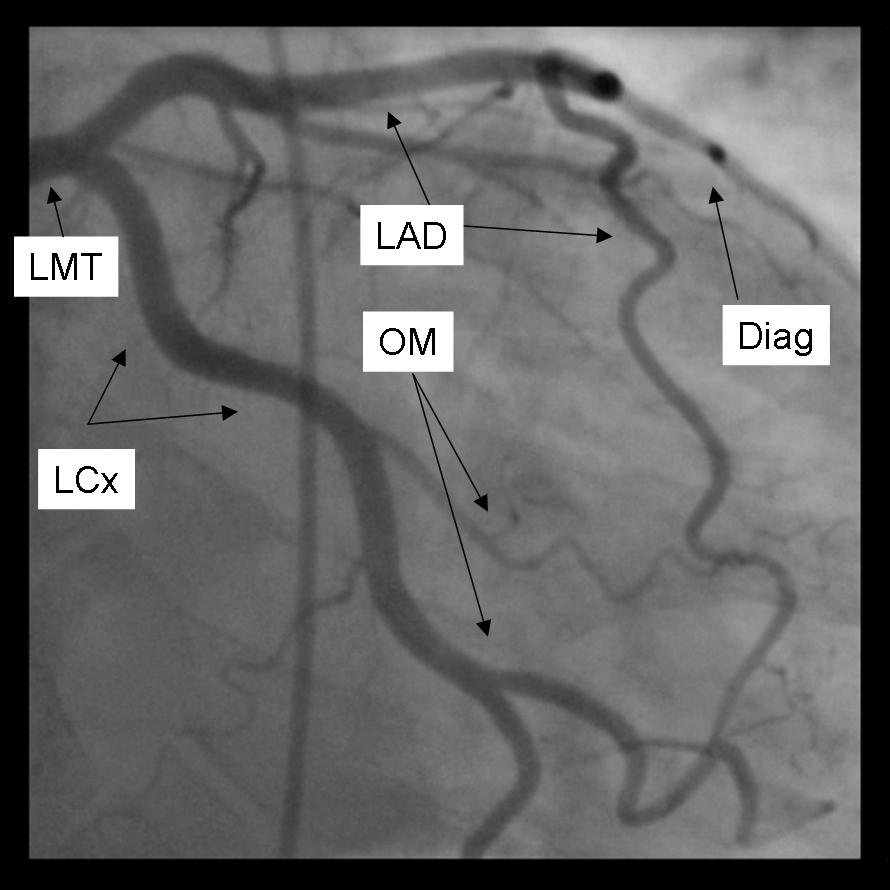
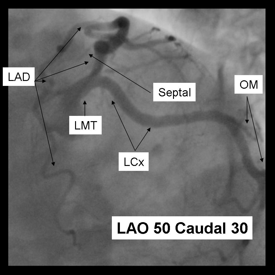
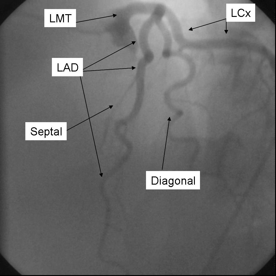
Additional images
-
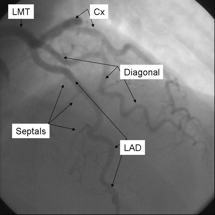
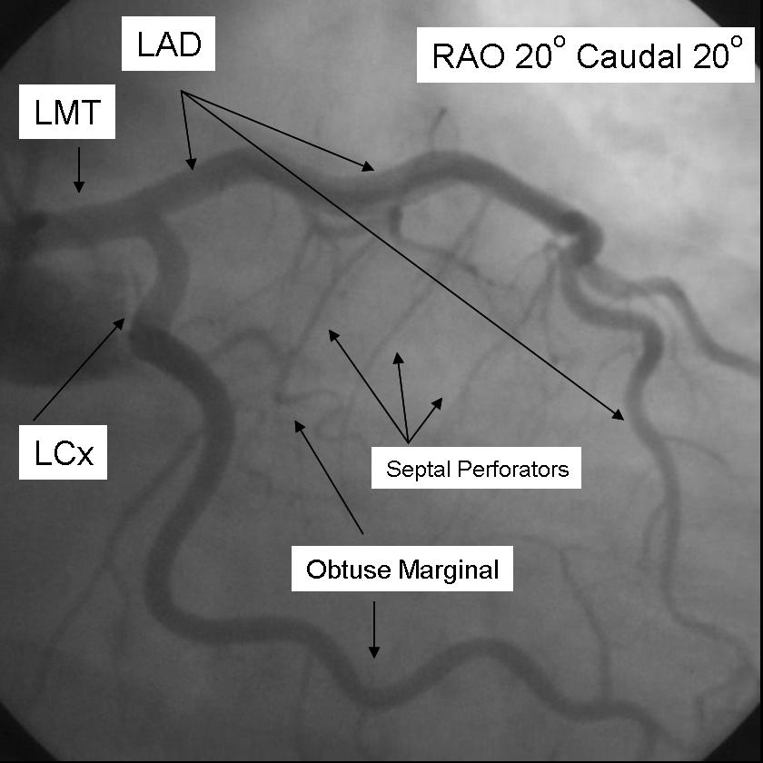 A coronary angiogram that shows the LMCA, LAD and LCX
 -
 A coronary angiogram that shows the LMCA, LAD and LCX
 -
 A coronary angiogram that shows the LMCA, LAD and LCX
 -
 A coronary angiogram that shows the LMCA, LAD and LCX
 -
 A coronary angiogram that shows the LMCA, LAD and LCX


