Restriction enzyme

Overview
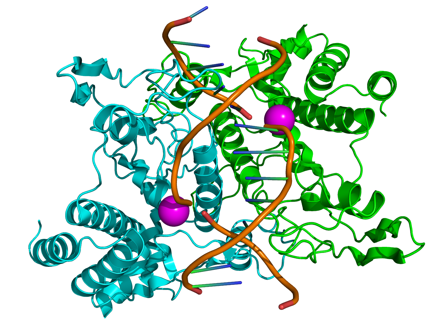
A restriction enzyme (or restriction endonuclease) is an enzyme that cuts double-stranded DNA. The enzyme makes two incisions, one through each of the sugar-phosphate backbones (i.e., each strand) of the double helix without damaging the nitrogenous bases. They work with cutting up foreign DNA, a process called restriction. Restriction enzymes therefore are believed to be a mechanism evolved by bacteria to resist attack from viruses called bacteriophages and to help in the removal of viral sequences. They are part of what is called the restriction modification system.
The chemical bonds that the enzymes cleave can be reformed by other enzymes known as ligases, so that restriction fragments carved from different chromosomes or genes can be spliced together, provided their ends are complementary (more below). Many of the procedures of molecular biology and genetic engineering rely on restriction enzymes.
The 1978 Nobel Prize in Medicine was awarded to the well-known scientist Daniel Nathans, Werner Arber and Hamilton Smith for the discovery of restriction endonucleases, leading to the development of recombinant DNA technology. The first practical use of their work was the manipulation of E. coli bacteria to produce human insulin for diabetics.
Fragment complementarity and splicing
Because recognition sequences and cleavage sites differ between restriction enzymes, the length and the exact sequence of a sticky-end "overhang", as well as whether it is the 5' end or the 3' end strand that overhangs, depends on which enzyme produced it. Base-pairing between overhangs with complementary sequences enables two fragments to be joined or "spliced" by a DNA ligase. A sticky-end fragment can be ligated not only to the fragment from which it was originally cleaved, but also to any other fragment with a compatible sticky end.The sticky end is also called a cohesive end or complementary end in some reference. If a restriction enzyme has a non-degenerate palindromic cleavage site, all ends that it produces are compatible. Ends produced by different enzymes may also be compatible.
Restriction enzymes as tools
- See the main article on restriction digests.
Recognition sequences typically are only four to twelve nucleotides long. Because there are only so many ways to arrange the four nucleotides--A,C,G and T--into a four or eight or twelve nucleotide sequence, recognition sequences tend to "crop up" by chance in any long sequence. Furthermore, restriction enzymes specific to hundreds of distinct sequences have been identified and synthesized for sale to laboratories. As a result, potential "restriction sites" appear in almost any gene or chromosome. Meanwhile, the sequences of some artificial plasmids include a "linker" that contains dozens of restriction enzyme recognition sequences within a very short segment of DNA. So no matter the context in which a gene naturally appears, there is probably a pair of restriction enzymes that can snip it out, and which will produce ends that enable the gene to be spliced into a "plasmid" (i.e. which will enable what molecular biologists call "cloning" of the gene).
Another use of restriction enzymes can be to find specific SNPs. If a restriction enzyme can be found such that it cuts only one possible allele of a section of DNA (that is, the alternate nucleotide of the SNP causes the restriction site to no longer exist within the section of DNA), this restriction enzyme can be used to genotype the sample without completely sequencing it. The sample is first run in a restriction digest to cut the DNA, then gel electrophoresis is performed on this digest. If the sample is homozygous for the common allele, the result will be two bands of DNA, because the cut will have occurred at the restriction site. If the sample is homozygous for the rarer allele, the sample will show only one band, because it will not have been cut. If the sample is heterozygous at that SNP, there will be three bands of DNA. This is an example of restriction mapping, see the article on restriction maps
Recognition sites
Restriction enzymes recognize a specific sequence of nucleotides and produce a double stranded cut in the DNA that prevents the phage from replicating. While recognition sequences vary widely, with lengths between 4 and 8 nucleotides, many of them are palindromic; that is, the sequence on one strand reads the same in the same direction on the complementary strand. The meaning of "palindromic" in this context is different from what one might expect from its linguistic usage: GTAATG is not a palindromic DNA sequence, but GTATAC is (GTATAC is complementary to CATATG).
Bacteria prevent their own DNA from being cut by modifying their nucleotides via methylation.
Different types of restriction enzymes
Restriction enzymes are of three types.i,2,3
Naming
Restriction enzymes are named based on the bacteria in which they are isolated in the following manner:
| E | Escherichia | (genus) |
| co | coli | (species) |
| R | RY13 | (strain) |
| I | First identified | Order ID'd in bacterium |
Enzyme Mechanisms
There are three groups of restriction enzyme that vary in the way recognise their restriction sites and where they cut the DNA:
- Type I restriction enzymes cut DNA about 100 nucleotides after the recognition site and requires ATP.
- Type II restriction enzymes cut DNA at the recognition site or near the recognition site (Type IIS) and for this reason are most often used in scientific experimentation.
- Type III restriction enzymes cut DNA about 20-30 base pairs after the recognition site and requires ATP.
Examples
| Enzyme | Source | Recognition Sequence | Cut |
|---|---|---|---|
| EcoRI | Escherichia coli |
5'GAATTC 3'CTTAAG |
5'---G AATTC---3' 3'---CTTAA G---5' |
| EcoRII | Escherichia coli |
5'CCWGG 3'GGWCC |
5'--- CCWGG---3' 3'---GGWCC ---5' |
| BamHI | Bacillus amyloliquefaciens |
5'GGATCC 3'CCTAGG |
5'---G GATCC---3' 3'---CCTAG G---5' |
| HindIII | Haemophilus influenzae |
5'AAGCTT 3'TTCGAA |
5'---A AGCTT---3' 3'---TTCGA A---5' |
| TaqI | Thermus aquaticus |
5'TCGA 3'AGCT |
5'---T CGA---3' 3'---AGC T---5' |
| NotI | Nocardia otitidis |
5'GCGGCCGC 3'CGCCGGCG |
5'---GC GGCCGC---3' 3'---CGCCGG CG---5' |
| HinfI | Haemophilus influenzae |
5'GANTC 3'CTNAG |
5'---G ANTC---3' 3'---CTNA G---5' |
| Sau3A | Staphylococcus aureus |
5'GATC 3'CTAG |
5'--- GATC---3' 3'---CTAG ---3' |
| PovII* | Proteus vulgaris |
5'CAGCTG 3'GTCGAC |
5'---CAG CTG---3' 3'---GTC GAC---5' |
| SmaI* | Serratia marcescens |
5'CCCGGG 3'GGGCCC |
5'---CCC GGG---3' 3'---GGG CCC---5' |
| HaeIII* | Haemophilus aegyptius |
5'GGCC 3'CCGG |
5'---GG CC---3' 3'---CC GG---5' |
| AluI* | Arthrobacter luteus |
5'AGCT 3'TCGA |
5'---AG CT---3' 3'---TC GA---5' |
| EcoRV* | Escherichia coli |
5'GATATC 3'CTATAG |
5'---GAT ATC---3' 3'---CTA TAG---5' |
| KpnI[1] | Klebsiella pneumoniae |
5'GGTACC 3'CCATGG |
5'---GGTAC C---3' 3'---C CATGG---5' |
| PstI[1] | Providencia stuartii |
5'CTGCAG 3'GACGTC |
5'---CTGCA G---3' 3'---G ACGTC---5' |
| SacI[1] | Streptomyces achromogenes |
5'GAGCTC 3'CTCGAG |
5'---GAGCT C---3' 3'---C TCGAG---5' |
| SalI[1] | Streptomyces albus |
5'GTCGAC 3'CAGCTG |
5'---G TCGAC---3' 3'---CAGCT G---5' |
| ScaI[1] | Streptomyces caespitosus |
5'AGTACT 3'TCATGA |
5'---AGT ACT---3' 3'---TCA TGA---5' |
| SphI[1] | Streptomyces phaeochromogenes |
5'GCATGC 3'CGTACG |
5'---G CATGC---3' 3'---CGTAC G---5' |
| StuI [2] | Streptomyces tubercidicus |
5'AGGCCT 3'TCCGGA |
5'---AGG CCT---3' 3'---TCC GGA---5' |
| XbaI[1] | Xanthomonas badrii |
5'TCTAGA 3'AGATCT |
5'---T CTAGA---3' 3'---AGATC T---5' |
| * = blunt ends | |||
| N = C or G or T or A | |||
| W = A or T | |||
See also
External links
- Restriction enzymes: protein data bank molecule of the month
- REBASE - The Restriction Enzyme Database
- Restriction enzyme finder
- pDRAW32 - Freeware software for restriction analysis
- WatCut - An online tool for restriction analysis
- Restriction digest of DNA - Online tool, free source code (PHP)
- Restriction Homepage - Six different tools
- Restriction Endonucleases: Molecular Scissors for Specifically Cutting DNA - a review from the Science Creative Quarterly
- Type I Restriction-Modification Systems - Home Page
- DNA Restriction Enzymes at the US National Library of Medicine Medical Subject Headings (MeSH)
References
- ↑ Jump up to: 1.0 1.1 1.2 1.3 1.4 1.5 1.6 Molecular cell biology. Lodish, Harvey F. 5. ed. : - New York : W. H. Freeman and Co., 2003, 973 s. b ill. ISBN: 0-7167-4366-3
- ↑ R8013 Stu I from Streptomyces tubercidicus buffered aqueous glycerol solution
bg:Рестрикционна ендонуклеаза ca:Enzim de restricció da:Restriktionsenzym de:Restriktionsenzym eo:Restriktaj enzimoj fa:اندونوکلئازهای محدودکننده it:Deossiribonucleasi II (sito-specifica) he:אנזים הגבלה nl:Restrictie-enzym fi:Restriktioentsyymi sv:Restriktionsenzym uk:Рестриктази