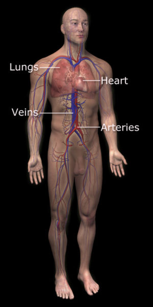
Cardiovascular system

Template:WikiDoc Cardiology News Editor-In-Chief: C. Michael Gibson, M.S., M.D. [1]
The circulatory system (or cardiovascular system) is an organ system that moves nutrients, gases, and wastes to and from cells, and helps stabilize body temperature and pH to maintain homeostasis. While humans, as well as other vertebrates have a closed circulatory system, some invertebrate groups have open circulatory system. The most primitive animal phyla lack circulatory systems.
Human circulatory system
The main components of the human circulatory system are the heart, the blood, and the blood vessels.
Furthermore, these components can either belong to the systemic circulation and the pulmonary circulation. The systemic circulation is the main part of the circulatory system, while the pulmonary system oxygenates the blood.
Systemic circulation
Systemic circulation is the portion of the cardiovascular system which carries oxygenated blood away from the heart, to the body, and returns deoxygenated blood back to the heart.
In the systemic circulation, arteries bring oxygenated blood to the tissues. As blood circulates through the body, oxygen diffuses from the blood into cells surrounding the capillaries, and carbon dioxide diffuses into the blood from the capillary cells. Veins bring deoxygenated blood back to the heart.
The release of oxygen from red blood cells or erythrocytes is regulated in mammals. It increases with an increase of carbon dioxide in tissues, an increase in temperature, or a decrease in pH. Such characteristics are exhibited by tissues undergoing high metabolism, as they require increased levels of oxygen.
Pulmonary circulation
Pulmonary circulation is the portion of the cardiovascular system which carries oxygen-depleted blood away from the heart, to the lungs, and returns oxygenated blood back to the heart.
De-oxygenated blood enters the right atrium of the heart and flows into the right ventricle where it is pumped through the pulmonary arteries to the lungs. Pulmonary veins return the now oxygen-rich blood to the heart, where it enters the left atrium before flowing into the left ventricle. From the left ventricle the oxygen-rich blood is pumped out via the aorta, and on to the rest of the body.
Heart
In the heart there is one atrium and one ventricle for each circulation, and with both a systemic and a pulmonary circulation there are four chambers in total: left atrium, left ventricle, right atrium and right ventricle.
Closed circulatory system
The circulatory systems of humans is closed, meaning that the blood never leaves the system of blood vessels. In contrast, oxygen and nutrients diffuse across the blood vessel layers and enters interstitial fluid, which carries oxygen and nutrients to the target cells, and carbon dioxide and wastes in the opposite direction.
Other vertebrates
The circulatory systems of all vertebrates, as well as of annelids (for example, earthworms) and cephalopods (squid and octopus) are closed, just as in humans. Still, the systems of fish, amphibians, reptiles, and birds show various stages of the evolution of the circulatory system.
In fish, the system has only one circuit, with the blood being pumped through the capillaries of the gills and on to the capillaries of the body tissues. This is known as single circulation. The heart of fish is therefore only a single pump (consisting of two chambers). In amphibians and most reptiles, a double circulatory system is used, but the heart is not always completely separated into two pumps. Amphibians have a three-chambered heart.
Birds and mammals show complete separation of the heart into two pumps, for a total of four heart chambers; it is thought that the four-chambered heart of birds evolved independently from that of mammals.
Open circulatory system
The open circulatory system is an arrangement of internal transport present in animals such as molluscs and arthropods, in which fluid (called hemolymph) in a cavity called the hemocoel bathes the organs directly and there is no distinction between blood and interstitial fluid; this combined fluid is called hemolymph or haemolymph. Muscular movements by the animal during locomotion can facilitate hemolymph movement, but diverting flow from one area to another is limited. When the heart relaxes, blood is drawn back toward the heart through open-ended pores.
Hemolymph fills all of the interior hemocoel of the body and surrounds all cells. Hemolymph is composed of water, inorganic salts (mostly Na+, Cl-, K+, Mg2+, and Ca2+), and organic compounds (mostly carbohydrates, proteins, and lipids). The primary oxygen transporter molecule is hemocyanin.
There are free-floating cells, the hemocytes, within the hemolymph. They play a role in the arthropod immune system.
No circulatory system
Circulatory systems are absent in some animals, including flatworms (phylum Platyhelminthes). Their body cavity has no lining or enclosed fluid. Instead a muscular pharynx leads to an extensively branched digestive system that facilitates direct diffusion of nutrients to all cells. The flatworm's dorso-ventrally flattened body shape also restricts the distance of any cell from the digestive system or the exterior of the organism. Oxygen can diffuse from the surrounding water into the cells, and carbon dioxide can diffuse out. Consequently every cell is able to obtain nutrients, water and oxygen without the need of a transport system.
Measurement techniques
Health and disease
History of discovery
The valves of the heart were discovered by a physician of the Hippocratean school around the 4th century BC. However their function was not properly understood then. Because blood pools in the veins after death, arteries look empty. Ancient anatomists assumed they were filled with air and that they were for transport of air.
Herophilus distinguished veins from arteries but thought that the pulse was a property of arteries themselves. Erasistratus observed that arteries that were cut during life bleed. He ascribed the fact to the phenomenon that air escaping from an artery is replaced with blood that entered by very small vessels between veins and arteries. Thus he apparently postulated capillaries but with reversed flow of blood.
The 2nd century AD Greek physician, Galen knew that blood vessels carried blood and identified venous (dark red) and arterial (brighter and thinner) blood, each with distinct and separate functions. Growth and energy were derived from venous blood created in the liver from chyle, while arterial blood gave vitality by containing pneuma (air) and originated in the heart. Blood flowed from both creating organs to all parts of the body where it was consumed and there was no return of blood to the heart or liver. The heart did not pump blood around, the heart's motion sucked blood in during diastole and the blood moved by the pulsation of the arteries themselves.
Galen believed that the arterial blood was created by venous blood passing from the left ventricle to the right by passing through 'pores' in the interventricular septum, air passed from the lungs via the pulmonary artery to the left side of the heart. As the arterial blood was created 'sooty' vapors were created and passed to the lungs also via the pulmonary artery to be exhaled.
In 1242 the Arab physician Ibn al-Nafis became the first person to accurately describe the process of blood circulation in the human body, including pulmonary circulation. He stated:
"...the blood from the right chamber of the heart must arrive at the left chamber but there is no direct pathway between them. The thick septum of the heart is not perforated and does not have visible pores as some people thought or invisible pores as Galen thought. The blood from the right chamber must flow through the vena arteriosa (pulmonary artery) to the lungs, spread through its substances, be mingled there with air, pass through the arteria venosa (pulmonary vein) to reach the left chamber of the heart and there form the vital spirit..."
Contemporary drawings of this process have survived. In 1552, Michael Servetus described the same, and Realdo Colombo proved the concept, but it remained largely unknown in Europe.
Finally William Harvey, a pupil of Hieronymus Fabricius (who had earlier described the valves of the veins without recognizing their function), performed a sequence of experiments and announced in 1628 the discovery of the human circulatory system as his own and published an influential book about it. This work with its essentially correct exposition slowly convinced the medical world. Harvey was not able to identify the capillary system connecting arteries and veins; these were later described by Marcello Malpighi.
See also
- Cardiology
- Lymphatic system
- Noise health effects
- Blood vessels
- Innate heat
- Cardiac muscle
- Major systems of the human body
- Heart
- Amato Lusitano
- William Harvey
References
- Iskandar, Albert Z. "Comprehensive Book on the Art of Medicine by Ibn al-Nafis". Retrieved May 2, 2005.
- Nie Jing-bao, " Refutation of the Claim that the Ancient Chinese described the Circulation of Blood," New Zealand Journal of Asian Studies 3, 2 (December, 2001): 119-135
External links
- The Circulatory System, a comprehensive overview
- The InVision Guide to a Healthy Heart An interactive website
- NCP Cardiovascular Medicine A Journal Covering Clinical Cardiovascular Medicine
Template:Development of circulatory system
ar:جهاز الدوران ast:Sistema cardiovascular bn:সংবহন তন্ত্র zh-min-nan:Sûn-khoân hē-thóng bs:Krvotok bg:Кръвообращение ca:Sistema cardiovascular cs:Oběhová soustava de:Blutkreislauf dv:ލޭދައުރުކުރާ ނިޒާމް eo:Kardiovaskula sistemo eu:Zirkulazio-aparatu gl:Aparello circulatorio hr:Krvožilni sustav id:Sistem kardiovaskular is:Blóðrásarkerfi it:Apparato circolatorio he:מחזור הדם pam:Circulatory system ku:Sîstema gera xwînê lv:Asinsrites orgānu sistēma lt:Kraujotakos sistema hu:Keringési rendszer mk:Циркулаторен систем nl:Hart- en vaatstelsel no:Sirkulasjonssystem nn:Kretsløpssystem qu:Sirk'a llika sq:Sistemi i qarkullimit të gjakut simple:Circulatory system sk:Obehová sústava sr:Крвни систем fi:Verenkierto sv:Kardiovaskulära systemet th:ระบบไหลเวียนโลหิต yi:בלוט צירקולאציע