Right bundle branch block
| Right bundle branch block | |
 | |
|---|---|
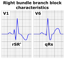
| ECG characteristics of a typical RBBB showing wide QRS complexes with a terminal R wave in lead V1 and slurred S wave in lead V6. |
|
Right bundle branch block Microchapters |
|
Differentiating Right bundle branch block from other Diseases |
|---|
|
Diagnosis |
|
Treatment |
|
Case Studies |
|
Right bundle branch block On the Web |
|
American Roentgen Ray Society Images of Right bundle branch block |
|
Risk calculators and risk factors for Right bundle branch block |
For patient information click here
Editor-In-Chief: C. Michael Gibson, M.S., M.D. [1]; Associate Editor(s)-in-Chief: Cafer Zorkun, M.D., Ph.D. [2]; Aarti Narayan, M.B.B.S [3]; Raviteja Guddeti, M.B.B.S. [4]
Synonyms and keywords: RBBB; bundle branch block right; rt bundle branch block
Overview
Historical Perspective
Classification
Pathophysiology
Causes
Differentiating Right bundle branch block from other Diseases
Epidemiology and Demographics
Risk Factors
Natural History, Complications and Prognosis
Diagnosis
History and Symptoms | Physical Examination | Laboratory Findings | Electrocardiogram | EKG Examples | Echocardiography | Other Imaging Findings | Other Diagnostic Studies
Treatment
Medical Therapy | Surgery | Primary Prevention | Secondary Prevention | Cost-Effectiveness of Therapy | Future or Investigational Therapies