Lymphadenopathy x ray

Revision as of 16:48, 8 March 2013 by Shankar Kumar (talk | contribs)
Jump to navigation Jump to search

Lymphadenopathy Microchapters

Home

Patient Information

Overview

Historical Perspective

Classification

Pathophysiology

Causes

Differentiating Lymphadenopathy from other Diseases

Epidemiology and Demographics

Risk Factors

Screening

Natural History, Complications and Prognosis

Diagnosis

Diagnostic Criteria

History and Symptoms

Physical Examination

Laboratory Findings

Electrocardiogram

X Ray

CT scan

MRI

Ultrasound

Other Imaging Findings

Other Diagnostic Studies

Treatment

Medical Therapy

Surgery

Primary Prevention

Secondary Prevention

Cost-Effectiveness of Therapy

Future or Investigational Therapies

Case Studies

Case #1

Lymphadenopathy x ray On the Web

Most recent articles

Most cited articles

Review articles

CME Programs

Powerpoint slides

Images

American Roentgen Ray Society Images of Lymphadenopathy x ray

All Images
X-rays
Echo & Ultrasound
CT Images
MRI

Ongoing Trials at Clinical Trials.gov

US National Guidelines Clearinghouse

NICE Guidance

FDA on Lymphadenopathy x ray

CDC on Lymphadenopathy x ray

Lymphadenopathy x ray in the news

Blogs on Lymphadenopathy x ray

Directions to Hospitals Treating Lymphadenopathy

Risk calculators and risk factors for Lymphadenopathy x ray

Editor-In-Chief: C. Michael Gibson, M.S., M.D. [1]; Associate Editor(s)-in-Chief: Raviteja Guddeti, M.B.B.S. [2]

Chest X Ray

Indications for chest x-ray in lymphadenopathy:

Bihilar lymphadenopathy is a radiographic term that describes the enlargement of mediastinal lymph nodes. It is easily and most commonly identified by a chest x-ray.
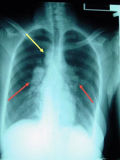
References

Template:WH Template:WS