(32 intermediate revisions by the same user not shown)
Line 7:
Line 7:
==Historical Perspective==
==Historical Perspective==
*In 1882, [[Neurofibromatosis 1|Neurofibromatosis]] ([[NF]]), described by Friedrich Daniel [[Von Recklinghausen neurofibromatosis|Von Recklinghausen]].<ref name="pmid23793209">{{cite journal| author=Antônio JR, Goloni-Bertollo EM, Trídico LA| title=Neurofibromatosis: chronological history and current issues. | journal=An Bras Dermatol | year= 2013 | volume= 88 | issue= 3 | pages= 329-43 | pmid=23793209 | doi=10.1590/abd1806-4841.20132125 | pmc=3754363 | url=https://www.ncbi.nlm.nih.gov/entrez/eutils/elink.fcgi?dbfrom=pubmed&tool=sumsearch.org/cite&retmode=ref&cmd=prlinks&id=23793209 }}</ref><ref name="Hosoi1931">{{cite journal|last1=Hosoi|first1=Kiyoshi|title=MULTIPLE NEUROFIBROMATOSIS (von RECKLINGHAUSEN'S DISEASE)|journal=Archives of Surgery|volume=22|issue=2|year=1931|pages=258|issn=0272-5533|doi=10.1001/archsurg.1931.01160020081004}}</ref>
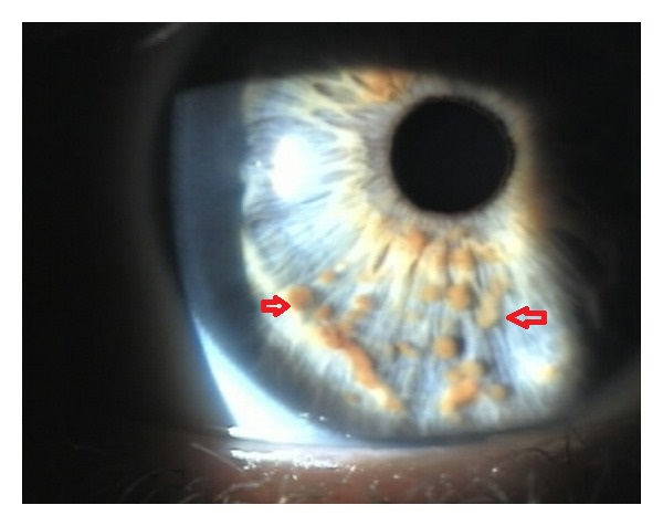
*[[File:Lisch Nodule.jpg|alt=Lisch nodules|thumb|Multiple small, oval, yellow-brown [[papules]] (Lisch nodules) in the right [[Iris (anatomy)|iris]](Red arrows). case courtesy by E. G. Adams et al.<ref>{{Cite web|url=https://www.ncbi.nlm.nih.gov/pmc/articles/PMC3350217/|title=Multiple, Unilateral Lisch Nodules in the Absence of Other Manifestations of Neurofibromatosis Type 1|last=|first=|date=|website=|archive-url=|archive-date=|dead-url=|access-date=}}</ref>]]In 1882, [[Neurofibromatosis 1|Neurofibromatosis]] ([[NF]]), described by Friedrich Daniel [[Von Recklinghausen neurofibromatosis|Von Recklinghausen]].<ref name="pmid23793209">{{cite journal| author=Antônio JR, Goloni-Bertollo EM, Trídico LA| title=Neurofibromatosis: chronological history and current issues. | journal=An Bras Dermatol | year= 2013 | volume= 88 | issue= 3 | pages= 329-43 | pmid=23793209 | doi=10.1590/abd1806-4841.20132125 | pmc=3754363 | url=https://www.ncbi.nlm.nih.gov/entrez/eutils/elink.fcgi?dbfrom=pubmed&tool=sumsearch.org/cite&retmode=ref&cmd=prlinks&id=23793209 }}</ref><ref name="Hosoi1931">{{cite journal|last1=Hosoi|first1=Kiyoshi|title=MULTIPLE NEUROFIBROMATOSIS (von RECKLINGHAUSEN'S DISEASE)|journal=Archives of Surgery|volume=22|issue=2|year=1931|pages=258|issn=0272-5533|doi=10.1001/archsurg.1931.01160020081004}}</ref>
*[[NF]] is a neuroectodermal abnormality constituted by a [[set]] of [[clinical]] [[symptoms]] that compromise the [[skin]], [[nervous system]], [[bones]], [[eyes]] and other sites.
*[[NF]] is a neuroectodermal abnormality constituted by a [[set]] of [[clinical]] [[symptoms]] that compromise the [[skin]], [[nervous system]], [[bones]], [[eyes]] and other sites.
*Lisch Nodules were named after the infamous Austrian [[ophthalmology|ophthalmologist]] Karl Lisch(1907-1999) who was also called as "Ophthalmological Pope".
*Lisch Nodules were named after the infamous Austrian [[ophthalmology|ophthalmologist]] Karl Lisch(1907-1999) who was also called as "Ophthalmological Pope".
*Besides general [[ophthalmology]], Lisch was interested in [[scientific]] [[research]]. He published more than 120 [[scientific]] papers in German [[Science (journal)|science]] journals.
*Besides general [[ophthalmology]], Lisch was interested in [[scientific]] [[research]]. He published more than 120 [[scientific]] papers in German [[Science (journal)|science]] journals.
*In 1937, Karl Lisch published an article on the [[iris]] [[hamartomas]] and their association with [[neurofibromatosis 1]], now known as "Lisch nodules", while at the University Eye Clinic in Munich.
*In 1937, Karl Lisch published an article on the [[iris]] [[hamartomas]] and their association with [[neurofibromatosis 1]], now known as "Lisch nodules", while at the University Eye Clinic in Munich.
*Lisch'a article described his [[Observation|observations]] in 3 [[patients]] with [[neurofibromatosis]]:<ref name="pmid20418991">{{cite journal| author=Gabhane SK, Kotwal MN, Bobhate SK| title=Segmental neurofibromatosis: a report of 3 cases. | journal=Indian J Dermatol | year= 2010 | volume= 55 | issue= 1 | pages= 105-8 | pmid=20418991 | doi=10.4103/0019-5154.60366 | pmc=2856359 | url=https://www.ncbi.nlm.nih.gov/entrez/eutils/elink.fcgi?dbfrom=pubmed&tool=sumsearch.org/cite&retmode=ref&cmd=prlinks&id=20418991 }}</ref>
*Lisch'a article described his [[Observation|observations]] in 3 [[patients]] with [[neurofibromatosis]]:<ref name="pmid20418991">{{cite journal| author=Gabhane SK, Kotwal MN, Bobhate SK| title=Segmental neurofibromatosis: a report of 3 cases. | journal=Indian J Dermatol | year= 2010 | volume= 55 | issue= 1 | pages= 105-8 | pmid=20418991 | doi=10.4103/0019-5154.60366 | pmc=2856359 | url=https://www.ncbi.nlm.nih.gov/entrez/eutils/elink.fcgi?dbfrom=pubmed&tool=sumsearch.org/cite&retmode=ref&cmd=prlinks&id=20418991 }}</ref><ref name="Dimitrova2009">{{cite journal|last1=Dimitrova|first1=Valentina|title=A CASE OF NEUROFIBROMATOSIS TYPE 1|journal=Journal of IMAB - Annual Proceeding (Scientific Papers)|volume=14, 1|issue=2008|year=2009|pages=63–67|issn=1312773X|doi=10.5272/jimab.14-1-2010.63}}</ref>
**[[Patient]] 1: A 39 [[year]] [[Old age|old]] [[male]] who had been affected with several [[nodules]] and [[Pigmented lesions|pigmented]] [[lesions]] on the [[skin]], typical of [[neurofibromatosis]], since the [[age]] of 15 [[Year|years]]. His mother and his sisters had a similar [[disorder]]. Lisch observed several [[brown]] [[nodules]] on the surface of the [[iris]]. The [[nodules]] could be seen even without the [[slit lamp]] due to the greyish-blue [[color]] of the [[iris]].
**[[Patient]] 1: A 39 [[year]] [[Old age|old]] [[male]] who had been affected with several [[nodules]] and [[Pigmented lesions|pigmented]] [[lesions]] on the [[skin]], typical of [[neurofibromatosis]], since the [[age]] of 15 [[Year|years]]. His mother and his sisters had a similar [[disorder]]. Lisch observed several [[brown]] [[nodules]] on the surface of the [[iris]]. The [[nodules]] could be seen even without the [[slit lamp]] due to the greyish-blue [[color]] of the [[iris]].
**[[Patient]] 2: 27 [[year]] [[Old age|old]] [[patient]] with similar [[cutaneous]] and [[iris]] [[lesions]] could be detected along with a [[family]] [[History and Physical examination|history]] of [[neurofibromatosis]]. In comparison to the first [[patient]] the [[iris]] [[nodules]] were much more pigmented.
**[[Patient]] 2: 27 [[year]] [[Old age|old]] [[patient]] with similar [[cutaneous]] and [[iris]] [[lesions]] could be detected along with a [[family]] [[History and Physical examination|history]] of [[neurofibromatosis]]. In comparison to the first [[patient]] the [[iris]] [[nodules]] were much more pigmented.
Line 22:
Line 22:
==Pathophysiology==
==Pathophysiology==
*A [[hamartoma]] is defined as a [[benign]] [[tumor]] or [[nodular]] [[growth]] that is composed of [[Proliferation|proliferating]] [[Maturation|mature]] [[histologically]] normal [[cells]] that normally reside at the affected [[tissue]].<ref name="pmid19516012">{{cite journal| author=Boley S, Sloan JL, Pemov A, Stewart DR| title=A quantitative assessment of the burden and distribution of Lisch nodules in adults with neurofibromatosis type 1. | journal=Invest Ophthalmol Vis Sci | year= 2009 | volume= 50 | issue= 11 | pages= 5035-43 | pmid=19516012 | doi=10.1167/iovs.09-3650 | pmc=2883270 | url=https://www.ncbi.nlm.nih.gov/entrez/eutils/elink.fcgi?dbfrom=pubmed&tool=sumsearch.org/cite&retmode=ref&cmd=prlinks&id=19516012 }}</ref><ref name="pmid19539839">{{cite journal| author=Boyd KP, Korf BR, Theos A| title=Neurofibromatosis type 1. | journal=J Am Acad Dermatol | year= 2009 | volume= 61 | issue= 1 | pages= 1-14; quiz 15-6 | pmid=19539839 | doi=10.1016/j.jaad.2008.12.051 | pmc=2716546 | url=https://www.ncbi.nlm.nih.gov/entrez/eutils/elink.fcgi?dbfrom=pubmed&tool=sumsearch.org/cite&retmode=ref&cmd=prlinks&id=19539839 }}</ref>
*A [[hamartoma]] is defined as a [[benign]] [[tumor]] or [[nodular]] [[growth]] that is composed of [[Proliferation|proliferating]] [[Maturation|mature]] [[histologically]] normal [[cells]] that normally reside at the affected [[tissue]].<ref name="pmid19650418">{{cite journal| author=Terzi YK, Oguzkan-Balci S, Anlar B, Aysun S, Guran S, Ayter S| title=Reproductive decisions after prenatal diagnosis in neurofibromatosis type 1: importance of genetic counseling. | journal=Genet Couns | year= 2009 | volume= 20 | issue= 2 | pages= 195-202 | pmid=19650418 | doi= | pmc= | url=https://www.ncbi.nlm.nih.gov/entrez/eutils/elink.fcgi?dbfrom=pubmed&tool=sumsearch.org/cite&retmode=ref&cmd=prlinks&id=19650418 }}</ref><ref name="pmid19516012">{{cite journal| author=Boley S, Sloan JL, Pemov A, Stewart DR| title=A quantitative assessment of the burden and distribution of Lisch nodules in adults with neurofibromatosis type 1. | journal=Invest Ophthalmol Vis Sci | year= 2009 | volume= 50 | issue= 11 | pages= 5035-43 | pmid=19516012 | doi=10.1167/iovs.09-3650 | pmc=2883270 | url=https://www.ncbi.nlm.nih.gov/entrez/eutils/elink.fcgi?dbfrom=pubmed&tool=sumsearch.org/cite&retmode=ref&cmd=prlinks&id=19516012 }}</ref><ref name="pmid19539839">{{cite journal| author=Boyd KP, Korf BR, Theos A| title=Neurofibromatosis type 1. | journal=J Am Acad Dermatol | year= 2009 | volume= 61 | issue= 1 | pages= 1-14; quiz 15-6 | pmid=19539839 | doi=10.1016/j.jaad.2008.12.051 | pmc=2716546 | url=https://www.ncbi.nlm.nih.gov/entrez/eutils/elink.fcgi?dbfrom=pubmed&tool=sumsearch.org/cite&retmode=ref&cmd=prlinks&id=19539839 }}</ref><ref name="TheosKorf2006">{{cite journal|last1=Theos|first1=Amy|last2=Korf|first2=Bruce R.|title=Pathophysiology of Neurofibromatosis Type 1|journal=Annals of Internal Medicine|volume=144|issue=11|year=2006|pages=842|issn=0003-4819|doi=10.7326/0003-4819-144-11-200606060-00010}}</ref>
*[[NF1]] is due to [[mutations]] in the [[NF1]] [[gene]], located at [[chromosome]] 17q11.2.
*[[NF1]] is due to [[mutations]] in the [[NF1]] [[gene]], located at [[chromosome]] 17q11.2.<ref name="pmid20422842">{{cite journal| author=Cohen R, Shuper A| title=[Developmental manifestation in children with neurofibromatosis type 1]. | journal=Harefuah | year= 2010 | volume= 149 | issue= 1 | pages= 49-52, 61 | pmid=20422842 | doi= | pmc= | url=https://www.ncbi.nlm.nih.gov/entrez/eutils/elink.fcgi?dbfrom=pubmed&tool=sumsearch.org/cite&retmode=ref&cmd=prlinks&id=20422842 }}</ref><ref name="pmid115721742">{{cite journal| author=de Goede-Bolder A, Cnossen MH, Dooijes D, van den Ouweland AM, Niermeijer MF| title=[From gene to disease; neurofibromatosis type 1]. | journal=Ned Tijdschr Geneeskd | year= 2001 | volume= 145 | issue= 36 | pages= 1736-8 | pmid=11572174 | doi= | pmc= | url=https://www.ncbi.nlm.nih.gov/entrez/eutils/elink.fcgi?dbfrom=pubmed&tool=sumsearch.org/cite&retmode=ref&cmd=prlinks&id=11572174 }}</ref>
*Lisch nodules are generally not present in central NF([[neurofibromatosis]])/NF([[neurofibromatosis]])-2.
*[[Neurofibromin]], the [[protein]] product encoded by the [[gene]], is expressed in many [[tissues]], including [[brain]], [[kidney]], [[spleen]], and [[thymus]].
*[[Neurofibromin]], the [[protein]] product encoded by the [[gene]], is expressed in many [[tissues]], including [[brain]], [[kidney]], [[spleen]], and [[thymus]].
*[[Mutations]] in the [[NF1]] [[gene]] result in loss of production or reduced [[Function (biology)|function]] of [[protein]]; this causes a wide spectrum of clinical findings, including [[NF1]]-associated [[Tumor|tumors]].<ref name="LubsBauer1991">{{cite journal|last1=Lubs|first1=Marie-Louise E.|last2=Bauer|first2=Mislen S.|last3=Formas|first3=Maria E.|last4=Djokic|first4=Borivoje|title=Lisch Nodules in Neurofibromatosis Type 1|journal=New England Journal of Medicine|volume=324|issue=18|year=1991|pages=1264–1266|issn=0028-4793|doi=10.1056/NEJM199105023241807}}</ref>
*[[Mutations]] in the [[NF1]] [[gene]] result in loss of production or reduced [[Function (biology)|function]] of [[protein]]; this causes a wide spectrum of clinical findings, including [[NF1]]-associated [[Tumor|tumors]].<ref name="LubsBauer1991">{{cite journal|last1=Lubs|first1=Marie-Louise E.|last2=Bauer|first2=Mislen S.|last3=Formas|first3=Maria E.|last4=Djokic|first4=Borivoje|title=Lisch Nodules in Neurofibromatosis Type 1|journal=New England Journal of Medicine|volume=324|issue=18|year=1991|pages=1264–1266|issn=0028-4793|doi=10.1056/NEJM199105023241807}}</ref><ref name="Dimitrova20092">{{cite journal|last1=Dimitrova|first1=Valentina|title=A CASE OF NEUROFIBROMATOSIS TYPE 1|journal=Journal of IMAB - Annual Proceeding (Scientific Papers)|volume=14, 1|issue=2008|year=2009|pages=63–67|issn=1312773X|doi=10.5272/jimab.14-1-2010.63}}</ref>
*[[Histopathology|Histopathologically]], Lisch nodules are composed of [[melanocytes]] and [[spindle cells]], usually concentrated on the superficial layers of the [[iris]] [[stroma]].<ref name="pmid28979620">{{cite journal| author=Abaloun Y, Ajhoun Y| title=[Lisch nodule in neurofibromatosis type 1]. | journal=Pan Afr Med J | year= 2017 | volume= 27 | issue= | pages= 218 | pmid=28979620 | doi=10.11604/pamj.2017.27.218.11517 | pmc=5622834 | url=https://www.ncbi.nlm.nih.gov/entrez/eutils/elink.fcgi?dbfrom=pubmed&tool=sumsearch.org/cite&retmode=ref&cmd=prlinks&id=28979620 }}</ref>
*[[Histopathology|Histopathologically]], Lisch nodules are composed of [[melanocytes]] and [[spindle cells]], usually concentrated on the superficial layers of the [[iris]] [[stroma]].<ref name="pmid28979620">{{cite journal| author=Abaloun Y, Ajhoun Y| title=[Lisch nodule in neurofibromatosis type 1]. | journal=Pan Afr Med J | year= 2017 | volume= 27 | issue= | pages= 218 | pmid=28979620 | doi=10.11604/pamj.2017.27.218.11517 | pmc=5622834 | url=https://www.ncbi.nlm.nih.gov/entrez/eutils/elink.fcgi?dbfrom=pubmed&tool=sumsearch.org/cite&retmode=ref&cmd=prlinks&id=28979620 }}</ref><ref name="pmid150961513">{{cite journal| author=Richetta A, Giustini S, Recupero SM, Pezza M, Carlomagno V, Amoruso G et al.| title=Lisch nodules of the iris in neurofibromatosis type 1. | journal=J Eur Acad Dermatol Venereol | year= 2004 | volume= 18 | issue= 3 | pages= 342-4 | pmid=15096151 | doi=10.1111/j.1468-3083.2004.00915.x | pmc= | url=https://www.ncbi.nlm.nih.gov/entrez/eutils/elink.fcgi?dbfrom=pubmed&tool=sumsearch.org/cite&retmode=ref&cmd=prlinks&id=15096151 }}</ref><ref name="RichettaGiustini2004">{{cite journal|last1=Richetta|first1=A|last2=Giustini|first2=S|last3=Recupero|first3=SM|last4=Pezza|first4=M|last5=Carlomagno|first5=V|last6=Amoruso|first6=G|last7=Calvieri|first7=S|title=Lisch nodules of the iris in neurofibromatosis type 1|journal=Journal of the European Academy of Dermatology and Venereology|volume=18|issue=3|year=2004|pages=342–344|issn=0926-9959|doi=10.1111/j.1468-3083.2004.00915.x}}</ref>
*The [[spindle cells]] are larger than the normal [[Iris (anatomy)|iris]] [[melanocytes]].
*The [[spindle cells]] are larger than the normal [[Iris (anatomy)|iris]] [[melanocytes]].
*[[Immunohistochemical]] studies show positive reaction against the following
*[[Immunohistochemical]] studies show positive reaction against the following
Line 45:
Line 46:
=== Incidence ===
=== Incidence ===
* The [[incidence]] of Lisch nodules is approximately 1 in 2600 to 3000 individuals worldwide.<ref name="LubsBauer19912">{{cite journal|last1=Lubs|first1=Marie-Louise E.|last2=Bauer|first2=Mislen S.|last3=Formas|first3=Maria E.|last4=Djokic|first4=Borivoje|title=Lisch Nodules in Neurofibromatosis Type 1|journal=New England Journal of Medicine|volume=324|issue=18|year=1991|pages=1264–1266|issn=0028-4793|doi=10.1056/NEJM199105023241807}}</ref>
* The [[incidence]] of Lisch nodules is approximately 1 in 2600 to 3000 individuals worldwide.<ref name="LubsBauer19912">{{cite journal|last1=Lubs|first1=Marie-Louise E.|last2=Bauer|first2=Mislen S.|last3=Formas|first3=Maria E.|last4=Djokic|first4=Borivoje|title=Lisch Nodules in Neurofibromatosis Type 1|journal=New England Journal of Medicine|volume=324|issue=18|year=1991|pages=1264–1266|issn=0028-4793|doi=10.1056/NEJM199105023241807}}</ref><ref name="pmid196504182">{{cite journal| author=Terzi YK, Oguzkan-Balci S, Anlar B, Aysun S, Guran S, Ayter S| title=Reproductive decisions after prenatal diagnosis in neurofibromatosis type 1: importance of genetic counseling. | journal=Genet Couns | year= 2009 | volume= 20 | issue= 2 | pages= 195-202 | pmid=19650418 | doi= | pmc= | url=https://www.ncbi.nlm.nih.gov/entrez/eutils/elink.fcgi?dbfrom=pubmed&tool=sumsearch.org/cite&retmode=ref&cmd=prlinks&id=19650418 }}</ref>
*Approximately one-half of the cases are [[familial]] ([[inherited]]).<ref name="pmid20082463">{{cite journal| author=Evans DG, Howard E, Giblin C, Clancy T, Spencer H, Huson SM et al.| title=Birth incidence and prevalence of tumor-prone syndromes: estimates from a UK family genetic register service. | journal=Am J Med Genet A | year= 2010 | volume= 152A | issue= 2 | pages= 327-32 | pmid=20082463 | doi=10.1002/ajmg.a.33139 | pmc= | url=https://www.ncbi.nlm.nih.gov/entrez/eutils/elink.fcgi?dbfrom=pubmed&tool=sumsearch.org/cite&retmode=ref&cmd=prlinks&id=20082463 }}</ref>
*Approximately one-half of the cases are [[familial]] ([[inherited]]).<ref name="pmid20082463">{{cite journal| author=Evans DG, Howard E, Giblin C, Clancy T, Spencer H, Huson SM et al.| title=Birth incidence and prevalence of tumor-prone syndromes: estimates from a UK family genetic register service. | journal=Am J Med Genet A | year= 2010 | volume= 152A | issue= 2 | pages= 327-32 | pmid=20082463 | doi=10.1002/ajmg.a.33139 | pmc= | url=https://www.ncbi.nlm.nih.gov/entrez/eutils/elink.fcgi?dbfrom=pubmed&tool=sumsearch.org/cite&retmode=ref&cmd=prlinks&id=20082463 }}</ref><ref name="pmid150961512">{{cite journal| author=Richetta A, Giustini S, Recupero SM, Pezza M, Carlomagno V, Amoruso G et al.| title=Lisch nodules of the iris in neurofibromatosis type 1. | journal=J Eur Acad Dermatol Venereol | year= 2004 | volume= 18 | issue= 3 | pages= 342-4 | pmid=15096151 | doi=10.1111/j.1468-3083.2004.00915.x | pmc= | url=https://www.ncbi.nlm.nih.gov/entrez/eutils/elink.fcgi?dbfrom=pubmed&tool=sumsearch.org/cite&retmode=ref&cmd=prlinks&id=15096151 }}</ref>
*<nowiki/>The remainder are the result of [[De novo|de novo (]]<nowiki/>sporadic) [[mutations]].
*<nowiki/>The remainder are the result of [[De novo|de novo (]]<nowiki/>sporadic) [[mutations]].
Line 55:
Line 56:
=== Age ===
=== Age ===
* Lisch nodules are predominantly visible in children usually after the age of six years.
* Lisch nodules are predominantly visible in children usually after the age of six years.<ref name="pmid204228422">{{cite journal| author=Cohen R, Shuper A| title=[Developmental manifestation in children with neurofibromatosis type 1]. | journal=Harefuah | year= 2010 | volume= 149 | issue= 1 | pages= 49-52, 61 | pmid=20422842 | doi= | pmc= | url=https://www.ncbi.nlm.nih.gov/entrez/eutils/elink.fcgi?dbfrom=pubmed&tool=sumsearch.org/cite&retmode=ref&cmd=prlinks&id=20422842 }}</ref>
*Lisch Nodule [[incidence]] in [[NF1]] increases with age and their prevalence raises by about 10% per year of life, up to age 9.
*Lisch Nodule [[incidence]] in [[NF1]] increases with age and their prevalence raises by about 10% per year of life, up to age 9.
Line 67:
Line 68:
==Screening==
==Screening==
*Lisch Nodules are not regularly [[Screening|screened]], they’re usually detected when other [[signs]] and [[Symptom|symptoms]] of [[neurofibromatosis]] appear.<ref name="LubsBauer19913">{{cite journal|last1=Lubs|first1=Marie-Louise E.|last2=Bauer|first2=Mislen S.|last3=Formas|first3=Maria E.|last4=Djokic|first4=Borivoje|title=Lisch Nodules in Neurofibromatosis Type 1|journal=New England Journal of Medicine|volume=324|issue=18|year=1991|pages=1264–1266|issn=0028-4793|doi=10.1056/NEJM199105023241807}}</ref>
* Lisch Nodules are not regularly [[Screening|screened]], they’re usually detected when other [[signs]] and [[Symptom|symptoms]] of [[neurofibromatosis]] appear.<ref name="LubsBauer19913">{{cite journal|last1=Lubs|first1=Marie-Louise E.|last2=Bauer|first2=Mislen S.|last3=Formas|first3=Maria E.|last4=Djokic|first4=Borivoje|title=Lisch Nodules in Neurofibromatosis Type 1|journal=New England Journal of Medicine|volume=324|issue=18|year=1991|pages=1264–1266|issn=0028-4793|doi=10.1056/NEJM199105023241807}}</ref>
*However, Lisch nodules are seen in 95% of children with [[NF1]] by age 20.<ref name="pmid3103673">{{cite journal| author=Huson S, Jones D, Beck L| title=Ophthalmic manifestations of neurofibromatosis. | journal=Br J Ophthalmol | year= 1987 | volume= 71 | issue= 3 | pages= 235-8 | pmid=3103673 | doi=10.1136/bjo.71.3.235 | pmc=1041127 | url=https://www.ncbi.nlm.nih.gov/entrez/eutils/elink.fcgi?dbfrom=pubmed&tool=sumsearch.org/cite&retmode=ref&cmd=prlinks&id=3103673 }}</ref>
*However, Lisch nodules are seen in 95% of children with [[NF1]] by age 20.<ref name="pmid3103673">{{cite journal| author=Huson S, Jones D, Beck L| title=Ophthalmic manifestations of neurofibromatosis. | journal=Br J Ophthalmol | year= 1987 | volume= 71 | issue= 3 | pages= 235-8 | pmid=3103673 | doi=10.1136/bjo.71.3.235 | pmc=1041127 | url=https://www.ncbi.nlm.nih.gov/entrez/eutils/elink.fcgi?dbfrom=pubmed&tool=sumsearch.org/cite&retmode=ref&cmd=prlinks&id=3103673 }}</ref>
Line 73:
Line 76:
=== Natural History ===
=== Natural History ===
* The symptoms of Lisch Nodules usually develop in the first decade of life, and mostly [[asymptomatic]].
=== Complications ===
=== Complications ===
Common [[complications]] of Lisch Nodules include:
* [Complication 1]
* Common [[complications]] of Lisch Nodules include:<ref name="pmid15096151">{{cite journal| author=Richetta A, Giustini S, Recupero SM, Pezza M, Carlomagno V, Amoruso G et al.| title=Lisch nodules of the iris in neurofibromatosis type 1. | journal=J Eur Acad Dermatol Venereol | year= 2004 | volume= 18 | issue= 3 | pages= 342-4 | pmid=15096151 | doi=10.1111/j.1468-3083.2004.00915.x | pmc= | url=https://www.ncbi.nlm.nih.gov/entrez/eutils/elink.fcgi?dbfrom=pubmed&tool=sumsearch.org/cite&retmode=ref&cmd=prlinks&id=15096151 }}</ref><ref name="Dimitrova20093">{{cite journal|last1=Dimitrova|first1=Valentina|title=A CASE OF NEUROFIBROMATOSIS TYPE 1|journal=Journal of IMAB - Annual Proceeding (Scientific Papers)|volume=14, 1|issue=2008|year=2009|pages=63–67|issn=1312773X|doi=10.5272/jimab.14-1-2010.63}}</ref>
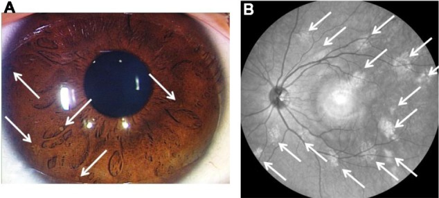
*[[File:Lisch nodules.jpg|alt=Lisch nodules and near-infrared reflectance image|thumb|Lisch nodules and near-infrared reflectance image (case 4). At least five Lisch nodules were detected and were classified as scale III (A). Note that 14 bright, patchy [[lesions]] were detected by near-infrared reflectance (B). The hyper-reflective point at the center of the image is an [[optical]] artifact. Case courtesy by Shinji Makino et al<ref>{{Cite web|url=https://www.ncbi.nlm.nih.gov/pmc/articles/PMC3883548/|title=Correlations between choroidal abnormalities, Lisch nodules, and age in patients with neurofibromatosis type 1|last=|first=|date=|website=|archive-url=|archive-date=|dead-url=|access-date=}}</ref>]]Lisch nodules occur in 90% of adults with [[Neurofibromatosis type I|neurofibromatosis]] 1.<ref name="pmid67892693">{{cite journal| author=Lewis RA, Riccardi VM| title=Von Recklinghausen neurofibromatosis. Incidence of iris hamartomata. | journal=Ophthalmology | year= 1981 | volume= 88 | issue= 4 | pages= 348-54 | pmid=6789269 | doi=10.1016/s0161-6420(81)35034-9 | pmc= | url=https://www.ncbi.nlm.nih.gov/entrez/eutils/elink.fcgi?dbfrom=pubmed&tool=sumsearch.org/cite&retmode=ref&cmd=prlinks&id=6789269 }}</ref><ref name="pmid31036733">{{cite journal| author=Huson S, Jones D, Beck L| title=Ophthalmic manifestations of neurofibromatosis. | journal=Br J Ophthalmol | year= 1987 | volume= 71 | issue= 3 | pages= 235-8 | pmid=3103673 | doi=10.1136/bjo.71.3.235 | pmc=1041127 | url=https://www.ncbi.nlm.nih.gov/entrez/eutils/elink.fcgi?dbfrom=pubmed&tool=sumsearch.org/cite&retmode=ref&cmd=prlinks&id=3103673 }}</ref><ref name="pmid289796203">{{cite journal| author=Abaloun Y, Ajhoun Y| title=[Lisch nodule in neurofibromatosis type 1]. | journal=Pan Afr Med J | year= 2017 | volume= 27 | issue= | pages= 218 | pmid=28979620 | doi=10.11604/pamj.2017.27.218.11517 | pmc=5622834 | url=https://www.ncbi.nlm.nih.gov/entrez/eutils/elink.fcgi?dbfrom=pubmed&tool=sumsearch.org/cite&retmode=ref&cmd=prlinks&id=28979620 }}</ref><ref name="pmid182803492">{{cite journal| author=Yang CC, Happle R, Chao SC, Yu-Yun Lee J, Chen W| title=Giant café-au-lait macule in neurofibromatosis 1: a type 2 segmental manifestation of neurofibromatosis 1? | journal=J Am Acad Dermatol | year= 2008 | volume= 58 | issue= 3 | pages= 493-7 | pmid=18280349 | doi=10.1016/j.jaad.2007.03.013 | pmc= | url=https://www.ncbi.nlm.nih.gov/entrez/eutils/elink.fcgi?dbfrom=pubmed&tool=sumsearch.org/cite&retmode=ref&cmd=prlinks&id=18280349 }}</ref><ref name="pmid204228424">{{cite journal| author=Cohen R, Shuper A| title=[Developmental manifestation in children with neurofibromatosis type 1]. | journal=Harefuah | year= 2010 | volume= 149 | issue= 1 | pages= 49-52, 61 | pmid=20422842 | doi= | pmc= | url=https://www.ncbi.nlm.nih.gov/entrez/eutils/elink.fcgi?dbfrom=pubmed&tool=sumsearch.org/cite&retmode=ref&cmd=prlinks&id=20422842 }}</ref><ref name="Dimitrova20094">{{cite journal|last1=Dimitrova|first1=Valentina|title=A CASE OF NEUROFIBROMATOSIS TYPE 1|journal=Journal of IMAB - Annual Proceeding (Scientific Papers)|volume=14, 1|issue=2008|year=2009|pages=63–67|issn=1312773X|doi=10.5272/jimab.14-1-2010.63}}</ref>
*[[Eye]]-findings include orange-brown colored specks.
*[[Eye]]-findings include orange-brown colored specks.
*Lisch nodules are usually elevated and tan in appearance.
*Lisch nodules are usually elevated and tan in appearance.
*Lisch nodules are [[benign]] [[hamartomas]] that can be seen without magnification.
*Lisch nodules are [[benign]] [[hamartomas]] that can be seen without magnification.
*Also known as [[Melanoma|melanocytic]] [[hamartomas]] of the iris, often associated with [[Neurofibromatosis type I|neurofibromatosis]] (NF) I.
*Also known as [[Melanoma|melanocytic]] [[hamartomas]] of the iris, often associated with [[Neurofibromatosis type I|neurofibromatosis]] (NF) I.<ref name="pmid14560838">{{cite journal| author=Nichols JC, Amato JE, Chung SM| title=Characteristics of Lisch nodules in patients with neurofibromatosis type 1. | journal=J Pediatr Ophthalmol Strabismus | year= 2003 | volume= 40 | issue= 5 | pages= 293-6 | pmid=14560838 | doi= | pmc= | url=https://www.ncbi.nlm.nih.gov/entrez/eutils/elink.fcgi?dbfrom=pubmed&tool=sumsearch.org/cite&retmode=ref&cmd=prlinks&id=14560838 }}</ref>
*Other associated [[Ophthalmology|ophthalmologic]] findings are [[Optic glioma|optic gliomas]].
*Other associated [[Ophthalmology|ophthalmologic]] findings are [[Optic glioma|optic gliomas]].
*[[Optic glioma|Optic gliomas]] can alter [[color vision]] and can produce progressive [[vision loss]].
*[[Optic glioma|Optic gliomas]] can alter [[color vision]] and can produce progressive [[vision loss]].
==Diagnostic Studies==
==Diagnostic Studies==
*On [[slit-lamp]] examination, they have a smooth, dome-shaped appearance and are usually light brown, although some can be very pale.<ref name="pmid67892692">{{cite journal| author=Lewis RA, Riccardi VM| title=Von Recklinghausen neurofibromatosis. Incidence of iris hamartomata. | journal=Ophthalmology | year= 1981 | volume= 88 | issue= 4 | pages= 348-54 | pmid=6789269 | doi=10.1016/s0161-6420(81)35034-9 | pmc= | url=https://www.ncbi.nlm.nih.gov/entrez/eutils/elink.fcgi?dbfrom=pubmed&tool=sumsearch.org/cite&retmode=ref&cmd=prlinks&id=6789269 }}</ref><ref name="pmid31036734">{{cite journal| author=Huson S, Jones D, Beck L| title=Ophthalmic manifestations of neurofibromatosis. | journal=Br J Ophthalmol | year= 1987 | volume= 71 | issue= 3 | pages= 235-8 | pmid=3103673 | doi=10.1136/bjo.71.3.235 | pmc=1041127 | url=https://www.ncbi.nlm.nih.gov/entrez/eutils/elink.fcgi?dbfrom=pubmed&tool=sumsearch.org/cite&retmode=ref&cmd=prlinks&id=3103673 }}</ref><ref name="pmid289796204">{{cite journal| author=Abaloun Y, Ajhoun Y| title=[Lisch nodule in neurofibromatosis type 1]. | journal=Pan Afr Med J | year= 2017 | volume= 27 | issue= | pages= 218 | pmid=28979620 | doi=10.11604/pamj.2017.27.218.11517 | pmc=5622834 | url=https://www.ncbi.nlm.nih.gov/entrez/eutils/elink.fcgi?dbfrom=pubmed&tool=sumsearch.org/cite&retmode=ref&cmd=prlinks&id=28979620 }}</ref>
*On [[slit-lamp]] examination, they have a smooth, dome-shaped appearance and are usually light brown, although some can be very pale.<ref name="pmid67892692">{{cite journal| author=Lewis RA, Riccardi VM| title=Von Recklinghausen neurofibromatosis. Incidence of iris hamartomata. | journal=Ophthalmology | year= 1981 | volume= 88 | issue= 4 | pages= 348-54 | pmid=6789269 | doi=10.1016/s0161-6420(81)35034-9 | pmc= | url=https://www.ncbi.nlm.nih.gov/entrez/eutils/elink.fcgi?dbfrom=pubmed&tool=sumsearch.org/cite&retmode=ref&cmd=prlinks&id=6789269 }}</ref><ref name="pmid31036734">{{cite journal| author=Huson S, Jones D, Beck L| title=Ophthalmic manifestations of neurofibromatosis. | journal=Br J Ophthalmol | year= 1987 | volume= 71 | issue= 3 | pages= 235-8 | pmid=3103673 | doi=10.1136/bjo.71.3.235 | pmc=1041127 | url=https://www.ncbi.nlm.nih.gov/entrez/eutils/elink.fcgi?dbfrom=pubmed&tool=sumsearch.org/cite&retmode=ref&cmd=prlinks&id=3103673 }}</ref><ref name="pmid289796204">{{cite journal| author=Abaloun Y, Ajhoun Y| title=[Lisch nodule in neurofibromatosis type 1]. | journal=Pan Afr Med J | year= 2017 | volume= 27 | issue= | pages= 218 | pmid=28979620 | doi=10.11604/pamj.2017.27.218.11517 | pmc=5622834 | url=https://www.ncbi.nlm.nih.gov/entrez/eutils/elink.fcgi?dbfrom=pubmed&tool=sumsearch.org/cite&retmode=ref&cmd=prlinks&id=28979620 }}</ref><ref name="pmid204228425">{{cite journal| author=Cohen R, Shuper A| title=[Developmental manifestation in children with neurofibromatosis type 1]. | journal=Harefuah | year= 2010 | volume= 149 | issue= 1 | pages= 49-52, 61 | pmid=20422842 | doi= | pmc= | url=https://www.ncbi.nlm.nih.gov/entrez/eutils/elink.fcgi?dbfrom=pubmed&tool=sumsearch.org/cite&retmode=ref&cmd=prlinks&id=20422842 }}</ref>
*[[Slit lamp]] examination can differentiate them from [[nevi]] on the [[iris]] by demonstrating elevated [[lesion]] instead of flat ones.
*[[Slit lamp]] examination can differentiate them from [[nevi]] on the [[iris]] by demonstrating elevated [[lesion]] instead of flat ones.<ref name="pmid19354164">{{cite journal| author=Crişan M, Talu S, Florea M, Coprean D, Cosgarea R, Crişan D| title=[Lisch nodules. Markers for a non-invasive diagnosis in Recklinghausen neurofibromatosis]. | journal=Oftalmologia | year= 2008 | volume= 52 | issue= 4 | pages= 56-61 | pmid=19354164 | doi= | pmc= | url=https://www.ncbi.nlm.nih.gov/entrez/eutils/elink.fcgi?dbfrom=pubmed&tool=sumsearch.org/cite&retmode=ref&cmd=prlinks&id=19354164 }}</ref>
*Lisch nodules develop during childhood, after the appearance of [[Café au lait spot|café-au-lait spots]] but before peripheral [[Neurofibroma|neurofibromas]].
*Lisch nodules develop during childhood, after the appearance of [[Café au lait spot|café-au-lait spots]] but before peripheral [[Neurofibroma|neurofibromas]].
*This is useful in confirming the diagnosis of [[NF1]] in children with no family history and only multiple [[Café-au-lait spot|café-au-lait spots]].
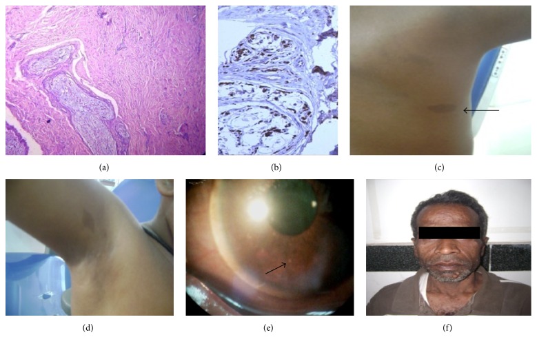
*[[File:Bundles of wavy spindle cells with serpentine nuclei .jpg|alt=Lisch nodule|thumb|(a) Microsection showing bundles of wavy [[spindle cells]] with serpentine nuclei in fascicles (H&E, ×10x), (b) Strong [[S-100 protein|S-100]] positivity of [[Tumor cell|tumor]] cells (×40x), (c) [[Café au lait spot|café au lait macule]] (arrow) in the back, (d) axillary [[freckle]], (e) Lisch nodule (arrow) in [[slit-lamp]] examination, and (f) father of patient with multiple cutaneous [[Neurofibroma|neurofibromas]]. Case courtesy by Rachna Rath et al.<ref>{{Cite web|url=https://www.ncbi.nlm.nih.gov/pmc/articles/PMC4921149/|title=Multifocal Head and Neck Neurofibromas with Osseous Abnormalities and Muscular Hypoplasia in a Child with Neurofibromatosis: Type I|last=|first=|date=|website=|archive-url=|archive-date=|dead-url=|access-date=}}</ref>]]This is useful in confirming the [[diagnosis]] of [[NF1]] in children with no [[family history]] and only multiple [[Café-au-lait spot|café-au-lait spots]].<ref name="pmid182803493">{{cite journal| author=Yang CC, Happle R, Chao SC, Yu-Yun Lee J, Chen W| title=Giant café-au-lait macule in neurofibromatosis 1: a type 2 segmental manifestation of neurofibromatosis 1? | journal=J Am Acad Dermatol | year= 2008 | volume= 58 | issue= 3 | pages= 493-7 | pmid=18280349 | doi=10.1016/j.jaad.2007.03.013 | pmc= | url=https://www.ncbi.nlm.nih.gov/entrez/eutils/elink.fcgi?dbfrom=pubmed&tool=sumsearch.org/cite&retmode=ref&cmd=prlinks&id=18280349 }}</ref><ref name="pmid196504184">{{cite journal| author=Terzi YK, Oguzkan-Balci S, Anlar B, Aysun S, Guran S, Ayter S| title=Reproductive decisions after prenatal diagnosis in neurofibromatosis type 1: importance of genetic counseling. | journal=Genet Couns | year= 2009 | volume= 20 | issue= 2 | pages= 195-202 | pmid=19650418 | doi= | pmc= | url=https://www.ncbi.nlm.nih.gov/entrez/eutils/elink.fcgi?dbfrom=pubmed&tool=sumsearch.org/cite&retmode=ref&cmd=prlinks&id=19650418 }}</ref>
==Treatment==
==Treatment==
Line 121:
Line 129:
=== Medical Therapy ===
=== Medical Therapy ===
*There is no treatment for the underlying disease nor any necessity to treat these small [[benign]] [[lesions]] which do not interfere with [[visual]] function.
*There is no treatment for the underlying disease nor any necessity to treat these small [[benign]] [[lesions]] which do not interfere with [[visual]] function.<ref name="Dimitrova20095">{{cite journal|last1=Dimitrova|first1=Valentina|title=A CASE OF NEUROFIBROMATOSIS TYPE 1|journal=Journal of IMAB - Annual Proceeding (Scientific Papers)|volume=14, 1|issue=2008|year=2009|pages=63–67|issn=1312773X|doi=10.5272/jimab.14-1-2010.63}}</ref>
*Lifelong monitoring is necessary because of the widespread manifestations and serious threat of [[complications]] such as:
*Lifelong monitoring is necessary because of the widespread manifestations and serious threat of [[complications]] such as:
**[[Visual]] [[impairment]]
**[[Visual]] [[impairment]]
**[[Renal]] [[hypertension]]
**[[Renal]] [[hypertension]]
**[[Ischemia]] of major [[Organ (anatomy)|organs]].
**[[Ischemia]] of major [[Organ (anatomy)|organs]].
== Primary Prevention ==
* There are no established measures for the [[primary prevention]] of Lisch nodules.
== Secondary Prevention ==
* There are no established measures for the [[secondary prevention]] of Lisch nodules.
In 1937, Karl Lisch published an article on the irishamartomas and their association with neurofibromatosis 1, now known as "Lisch nodules", while at the University Eye Clinic in Munich.
Lisch Nodules commonly associated with neurofibromatosis and is caused by genetic defects or mutations that either are passed on by a parent or occur spontaneously at conception.
The diagnosis is primarily based on clinical assessment and two or more of the features are required to confirm the diagnosis.
Physical Examination
Lisch nodules and near-infrared reflectance image (case 4). At least five Lisch nodules were detected and were classified as scale III (A). Note that 14 bright, patchy lesions were detected by near-infrared reflectance (B). The hyper-reflective point at the center of the image is an optical artifact. Case courtesy by Shinji Makino et al[38]Lisch nodules occur in 90% of adults with neurofibromatosis 1.[39][40][41][42][43][44]
↑Dimitrova, Valentina (2009). "A CASE OF NEUROFIBROMATOSIS TYPE 1". Journal of IMAB - Annual Proceeding (Scientific Papers). 14, 1 (2008): 63–67. doi:10.5272/jimab.14-1-2010.63. ISSN1312-773X.
↑Lubs, Marie-Louise E.; Bauer, Mislen S.; Formas, Maria E.; Djokic, Borivoje (1991). "Lisch Nodules in Neurofibromatosis Type 1". New England Journal of Medicine. 324 (18): 1264–1266. doi:10.1056/NEJM199105023241807. ISSN0028-4793.
↑Dimitrova, Valentina (2009). "A CASE OF NEUROFIBROMATOSIS TYPE 1". Journal of IMAB - Annual Proceeding (Scientific Papers). 14, 1 (2008): 63–67. doi:10.5272/jimab.14-1-2010.63. ISSN1312-773X.
↑Richetta, A; Giustini, S; Recupero, SM; Pezza, M; Carlomagno, V; Amoruso, G; Calvieri, S (2004). "Lisch nodules of the iris in neurofibromatosis type 1". Journal of the European Academy of Dermatology and Venereology. 18 (3): 342–344. doi:10.1111/j.1468-3083.2004.00915.x. ISSN0926-9959.
↑Lubs, Marie-Louise E.; Bauer, Mislen S.; Formas, Maria E.; Djokic, Borivoje (1991). "Lisch Nodules in Neurofibromatosis Type 1". New England Journal of Medicine. 324 (18): 1264–1266. doi:10.1056/NEJM199105023241807. ISSN0028-4793.
↑Lubs, Marie-Louise E.; Bauer, Mislen S.; Formas, Maria E.; Djokic, Borivoje (1991). "Lisch Nodules in Neurofibromatosis Type 1". New England Journal of Medicine. 324 (18): 1264–1266. doi:10.1056/NEJM199105023241807. ISSN0028-4793.
↑Dimitrova, Valentina (2009). "A CASE OF NEUROFIBROMATOSIS TYPE 1". Journal of IMAB - Annual Proceeding (Scientific Papers). 14, 1 (2008): 63–67. doi:10.5272/jimab.14-1-2010.63. ISSN1312-773X.
↑Dimitrova, Valentina (2009). "A CASE OF NEUROFIBROMATOSIS TYPE 1". Journal of IMAB - Annual Proceeding (Scientific Papers). 14, 1 (2008): 63–67. doi:10.5272/jimab.14-1-2010.63. ISSN1312-773X.
↑Dimitrova, Valentina (2009). "A CASE OF NEUROFIBROMATOSIS TYPE 1". Journal of IMAB - Annual Proceeding (Scientific Papers). 14, 1 (2008): 63–67. doi:10.5272/jimab.14-1-2010.63. ISSN1312-773X.