Right-to-left shunt
| Right-to-left shunt | |
 | |
|---|---|
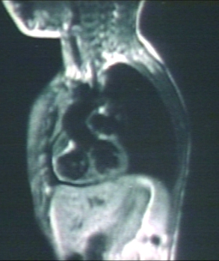
| MRI: Tetralogy of Fallot; Left anterior oblique view. with VSD and overriding aorta. Image courtesy of Professor Peter Anderson DVM PhD and published with permission © PEIR, University of Alabama at Birmingham, Department of Pathology |
Editor-In-Chief: C. Michael Gibson, M.S., M.D. [1]
Overview
A right-to-left shunt is a cardiac shunt which allows, or is designed to cause, blood to flow from the right heart to the left heart. This terminology is used both for the abnormal state in humans and for normal physiological shunts in reptiles. A right-to-left shunt occurs when there is an opening or passage between the atria, ventricles, and/or great vessels. For it to be considered a right to left shunt, the right heart pressure has to be higher than the left heart pressure and/or the shunt has a one-way valvular opening.
Causes
Common Causes
The most common cause of right-to-left shunt is the Tetralogy of Fallot, a congenital cardiac anomaly characterized by four co-existing heart defects. The four defects include:
- Pulmonary stenosis (narrowing of the pulmonary valve and outflow tract, obstructing blood flow from the right ventricle to the pulmonary artery)
- Ventricular septal defect (defect in the ventricular septum, which divides the left and right ventricles of the heart)
- Overriding aorta (aortic valve is enlarged and appears to arise from both the left and right ventricles instead of the left ventricle, as occurs in normal hearts)
- Right ventricular hypertrophy (thickening of the muscular walls of the right ventricle)
- Ebstein's anomaly
Natural History, Complications and Prognosis
A frequent complication resulting from a right to left shunt is hypoxemia.
References