[[File: Pathophysiology of WPW- Pre-excitation syndrome.jpg|thumb|Pathophysiology of WPW / Pre-excitation syndrome.[https://www.slideshare.net/smcmedicinedept/ecg-wpw-syndrome?next_slideshow=3]]]
[[File: Pathophysiology of WPW- Pre-excitation syndrome.jpg|thumb|Pathophysiology of WPW / Pre-excitation syndrome.[https://www.slideshare.net/smcmedicinedept/ecg-wpw-syndrome?next_slideshow=3]]]
* Normally the [[electrical]] activity in the [[heart]] starts from [[SA node]].
*Normally the [[electrical]] activity in the [[heart]] starts from [[SA node]].
* The [[impulse]] generation usually happens in the right [[atrium]] near the [[entrance]] of [[superior vena cava]] and it travels from [[SA node]] to the [[AV node]].<ref name="urlWolff-Parkinson-White pattern - Conditions - GTR - NCBI">{{cite web |url=https://www.ncbi.nlm.nih.gov/gtr/conditions/C0043202/ |title=Wolff-Parkinson-White pattern - Conditions - GTR - NCBI |format= |work= |accessdate=}}</ref>
*The [[impulse]] generation usually happens in the right [[atrium]] near the [[entrance]] of [[superior vena cava]] and it travels from [[SA node]] to the [[AV node]].
* The [[AV node ]] modulates the rate and number of [impulses]] to be conducted to the [[ventricles]]. The [[AV node]] also modulates the speed of transmission from [[atria]] to [[ventricles]] represents the [[PR interval]] on ECG. From the [[AV node]], an [[electrical]] [[impulse]] is transmitted to the [[bundle of His]], to left and right branches extending to the [[ventricular]] [[myocardium]].
*The [[AV node ]] modulates the rate and number of impulses to be conducted to the [[ventricles]].
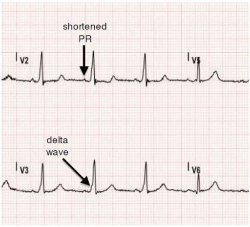
*The [[AV node]] also modulates the speed of transmission from [[atria]] to [[ventricles]] which represents the [[PR interval]] on ECG.
*From the [[AV node]], an [[electrical]] [[impulse]] is transmitted to the [[bundle of His]], to left and right branches extending to the [[ventricular]] [[myocardium]].
[[WPW]] is another word for [[pre-excitation]] of the [[ventricle]] through the [[accessory]] [[pathway]] instead of going through the usual pathway of [[AV node]] which usually slows down the [[speed]] of [[conduction]] of [[impulses]] transmitting to [[ventricles]]. The [[accessory]] pathway creates a channel directly to [[conduct]] the [[impulses]] to [[ventricles]] resulting in [[premature]] [[excitation]]. In "Type A [[Pre-excitation]]" [[accessory]] pathway lies between [[Left atria]] [[ventricles]] and in Type B [[pre-excitation]] fibers carry impulses between [[right atria]] and [[ventricles]].
* [[WPW]] is another word for [[pre-excitation]] of the [[ventricle]] through the [[accessory]] [[pathway]] instead of going through the usual pathway of [[AV node]] which usually slows down the [[speed]] of [[conduction]] of [[impulses]] transmitting to [[ventricles]].
* The [[accessory]] pathway creates a channel directly to [[conduct]] the [[impulses]] to [[ventricles]] resulting in [[premature]] [[excitation]].
* In "Type A [[Pre-excitation]]" [[accessory]] pathway lies between [[Left atria]] [[ventricles]] and in Type B [[pre-excitation]] fibers carry impulses between [[right atria]] and [[ventricles]].
Basic concept of Pathophysiology in [[pre-excitation syndrome]] lies in the concept of bypassing the [[AV node]] [[conduction]] and letting the [[impulse conduct]] faster through [[atria]] to [[ventricles]] via [[accessory pathways]]. These [[accessory pathways]] Usually called [[Bundle of Kent]] in [[WPW syndrome]], [[James fiber]] in [[LGL syndrome]] and [[Mahaim fibers]] in Mahaim type [[pre-excitation syndrome]]. These conducts [[impulses]] in forward (not common), backward ( around 15-20%) and in both directions ( Most common type) as well.
* Basic concept of pathophysiology in [[pre-excitation syndrome]] lies in the concept of bypassing the [[AV node]] [[conduction]] and letting the [[impulse conduct]] faster through [[atria]] to [[ventricles]] via [[accessory pathways]].
* These [[accessory pathways]] usually called [[Bundle of Kent]] in [[WPW syndrome]], [[James fiber]] in [[LGL syndrome]] and [[Mahaim fibers]] in Mahaim type [[pre-excitation syndrome]].
* These conducts [[impulses]] in forward (not common), backward ( around 15-20%) and in both directions (most common type) as well.
The [[accessory pathways]] mediate the occurrence of [[tachyarrhythmia]] by forming a [[re-entry]] circuit and commonly known as [[AVRT]]. The direct [[conduction]] of [[impulses]] from [[atria]] to [[ventricles]] can also result in the development of [[tachyarrhythmia's]] when there is a development of [[Atrial Fibrillation]] with [[RVR]]
* The [[accessory pathways]] mediate the occurrence of [[tachyarrhythmia]] by forming a [[re-entry]] circuit and commonly known as [[AVRT]].
* The direct [[conduction]] of [[impulses]] from [[atria]] to [[ventricles]] can also result in the development of [[tachyarrhythmia's]] when there is a development of [[Atrial Fibrillation]] with [[RVR]].
[[WPW syndrome]] is a combination of [[WPW]] pattern on [[ECG]] + [[Paroxysmal arrhythmias]]. The [[accessory pathways]] are usually named as [[Bundle of Kent]] or [[AV]] [[bypass tracts]]. [[Accessory pathways|The accessory pathways]] here are named as [[James fibers]], also known as [[Atrionodal fibers]] connecting the [[Atrium (heart)|atrium]] to the distal [[Atrioventricular node|AV node]]. These usually [[conduct]] the [[impulses]] from [[atria]] to the initial portion of the [[AV node]]. [[Accessory pathways|The accessory pathways]] named as [[Mahaim fibers]] connect the [[Atrium (heart)|Atrium]], [[AV node]], or [[bundle of His]] to the [[Purkinje fibers]] or [[ventricular myocardium]]. <br />
* [[WPW syndrome]] is a combination of [[WPW]] pattern on [[ECG]] + [[Paroxysmal arrhythmias]].
* The [[accessory pathways]] are usually named as [[Bundle of Kent]] or [[AV]] [[bypass tracts]].
* [[Accessory pathways|The accessory pathways]] here are named as [[James fibers]], also known as [[Atrionodal fibers]] connecting the [[Atrium (heart)|atrium]] to the distal [[Atrioventricular node|AV node]].
* These usually [[conduct]] the [[impulses]] from [[atria]] to the initial portion of the [[AV node]].
* [[Accessory pathways|The accessory pathways]] named as [[Mahaim fibers]] connect the [[Atrium (heart)|Atrium]], [[AV node]], or [[bundle of His]] to the [[Purkinje fibers]] or [[ventricular myocardium]]. <br />
==Differentiating Pre-excitation Syndrome from other Diseases==
==Differentiating Pre-excitation Syndrome from other Diseases==
Line 341:
Line 352:
===Natural History===
===Natural History===
*There are a lot of [[studies]] being done in the [[past]] to [[describe]] the [[natural history]] or [[disease]] course of [[pre-excitation syndrome]]. But data from a recent study- "[[Long term]] [[natural]] [[history]] of [[patients]] with [[WPW]] treated with or without [[catheter ablation]]" showed promising [[results]] in explaining the reduced [[long-term]] [[mortality]] rates in [[WPW]] patients who are matched for [[age]] and [[gender]]. Also explained the lower [[mortality]] rates in [[catheter]] ablated [[patients]] as compared to non ablated ones. Although the [[patients]] can die with [[sudden cardiac death]] but the rate of this scenaio is very less and not commonly observed<ref name="pmid22532593">{{cite journal |vauthors=Obeyesekere MN, Leong-Sit P, Massel D, Manlucu J, Modi S, Krahn AD, Skanes AC, Yee R, Gula LJ, Klein GJ |title=Risk of arrhythmia and sudden death in patients with asymptomatic preexcitation: a meta-analysis |journal=Circulation |volume=125 |issue=19 |pages=2308–15 |date=May 2012 |pmid=22532593 |doi=10.1161/CIRCULATIONAHA.111.055350 |url=}}</ref>.
*There are a lot of [[studies]] being done in the [[past]] to [[describe]] the [[natural history]] or [[disease]] course of [[pre-excitation syndrome]]. But data from a recent study- "[[Long term]] [[natural]] [[history]] of [[patients]] with [[WPW]] treated with or without [[catheter ablation]]" showed promising [[results]] in explaining the reduced [[long-term]] [[mortality]] rates in [[WPW]] patients who are matched for [[age]] and [[gender]]. Also explained the lower [[mortality]] rates in [[catheter]] ablated [[patients]] as compared to non ablated ones. Although the [[patients]] can die with [[sudden cardiac death]] but the rate of this scenaio is very less and not commonly observed.
===Complications===
===Complications===
Line 442:
Line 453:
[[File: Catheter Ablation.png|thumb|Catheter Ablation- Surgical Approach in WPW. Image showing catheter ablation of right free wall accessory pathway. The first successful ablation was performed by Morady and Scheinman. [https://openi.nlm.nih.gov/detailedresult?img=PMC3678820_rmmj-3-3-e0019_Figure5&query=WPW%20syndrome&it=xg&req=4&npos=49]]]
[[File: Catheter Ablation.png|thumb|Catheter Ablation- Surgical Approach in WPW. Image showing catheter ablation of right free wall accessory pathway. The first successful ablation was performed by Morady and Scheinman. [https://openi.nlm.nih.gov/detailedresult?img=PMC3678820_rmmj-3-3-e0019_Figure5&query=WPW%20syndrome&it=xg&req=4&npos=49]]]
<u>RADIOFREQUENCY ABLATION<ref name="pmid22579340">{{cite journal |vauthors=Cohen MI, Triedman JK, Cannon BC, Davis AM, Drago F, Janousek J, Klein GJ, Law IH, Morady FJ, Paul T, Perry JC, Sanatani S, Tanel RE |title=PACES/HRS expert consensus statement on the management of the asymptomatic young patient with a Wolff-Parkinson-White (WPW, ventricular preexcitation) electrocardiographic pattern: developed in partnership between the Pediatric and Congenital Electrophysiology Society (PACES) and the Heart Rhythm Society (HRS). Endorsed by the governing bodies of PACES, HRS, the American College of Cardiology Foundation (ACCF), the American Heart Association (AHA), the American Academy of Pediatrics (AAP), and the Canadian Heart Rhythm Society (CHRS) |journal=Heart Rhythm |volume=9 |issue=6 |pages=1006–24 |date=June 2012 |pmid=22579340 |doi=10.1016/j.hrthm.2012.03.050 |url=}}</ref></u>
<u>RADIOFREQUENCY ABLATION</u>
*This modality has replaced [[drug therapy]] and other [[Surgery operation|surgical treatment]] options by showing promising results. Best results are studied these [[days]] when it is used in [[Conjunction introduction|conjunction]] with [[cryoblation]] (commonly used for [[Accessory pathway|septal Accessory pathways]] and for [[accessory pathways]] near small [[coronary arteries]])
*This modality has replaced [[drug therapy]] and other [[Surgery operation|surgical treatment]] options by showing promising results. Best results are studied these [[days]] when it is used in [[Conjunction introduction|conjunction]] with [[cryoblation]] (commonly used for [[Accessory pathway|septal Accessory pathways]] and for [[accessory pathways]] near small [[coronary arteries]])
British physiologist "Albert Frank Stanley Kent" (1863 - 1958), first described the lateral branches of AV grove of the monkeyheart, which was later named accessory bundle of Kent.
Initial R wave in V1, initial r > 40 ms in V1/V2, notched S in V1, initial R in aVR, lead II R wave peak time ≥50 ms, no RS in V1-V6, and atrioventricular dissociation
WPW can be considered as a congenital anomaly in some cases where it is usually present since birth and in others and it is regarded as a developmental anomaly. Studies proved it's lowerprevalence in childrenaged between 6-13 than those in the age group of 14-15 years of age.
Catheter Ablation- Surgical Approach in WPW. Image showing catheter ablation of right free wall accessory pathway. The first successful ablation was performed by Morady and Scheinman. [3]
Patients who are not willing to undergo radiofrequencyablation can be managed on medical management with the use of Anti-arrhythmic. Though its role in the prevention of future episodes of arrhythmias is limited still this is the most commonly used modality of choice.


![[1]](https://en.ecgpedia.org/index.php?title=File:Wolffparkinsonwhite.jpg)

