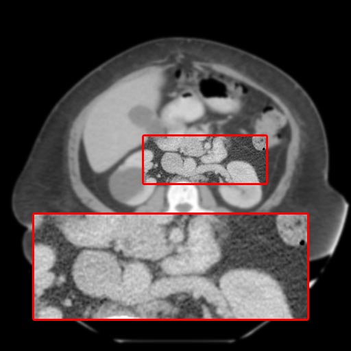
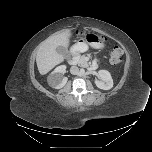
Retroaortic left renal vein
Jump to navigation
Jump to search
| Retroaortic left renal vein | |
 |
|---|
Editor-In-Chief: C. Michael Gibson, M.S., M.D. [1]
Associate Editor-In-Chief: Cafer Zorkun, M.D., Ph.D. [2]
Overview
- Seen in 2-3% of individuals (less common than circumaortic left renal vein, seen in 5-7%)
- Retroaortic left renal vein derived from the renal collar (circumaortic renal venous ring) at an embryonic stage and completed by the regression and disappearance of the ventral (preaortic) limb and the persistence of the dorsal (postaortic) limb at a later stage
- Drainage of lumbar veins may also be anomalous in these cases, although by variable anatomy
- In some cases, the retroaortic left renal vein exits the renal hilum posterior to the left renal artery (anomalous)
- The retroaortic vein may receive flow from lumbar veins and may run caudad and enter the inferior vena cava in the lower lumbar region, or it may subdivide before entering the inferior vena cava. [1] [2]
Diagnosis
Multi Sliced CT
Images shown below are courtesy of RadsWiki and copylefted
References
- ↑ Izumiyama, M, and Horiguchi, M. Two cases of the retroaortic left renal vein and a morphogenetic consideration of the anomalous vein. Kaibogaku Zasshi, 1997; 72(6): 535-543.
- ↑ Kawamoto, S, Montgomery, R.A., Lawler, L.P., Horton, K.M., and Fishman, E.K. Multi-detector row CT evaluation of living renal donors prior to laparoscopic nephrectomy. Radiographics, 2004; 24:453-466.
Additional Reading
- Moss and Adams' Heart Disease in Infants, Children, and Adolescents Hugh D. Allen, Arthur J. Moss, David J. Driscoll, Forrest H. Adams, Timothy F. Feltes, Robert E. Shaddy, 2007 ISBN 0781786843
