Left ventriculography Left Ventricular Ejection Fraction Assessment by Visual Estimation
|
Coronary Angiography | |
|
General Principles | |
|---|---|
|
Anatomy & Projection Angles | |
|
Normal Anatomy | |
|
Anatomic Variants | |
|
Projection Angles | |
|
Epicardial Flow & Myocardial Perfusion | |
|
Epicardial Flow | |
|
Myocardial Perfusion | |
|
Lesion Complexity | |
|
ACC/AHA Lesion-Specific Classification of the Primary Target Stenosis | |
|
Lesion Morphology | |
Editor-In-Chief: C. Michael Gibson, M.S., M.D. [1]
Overview
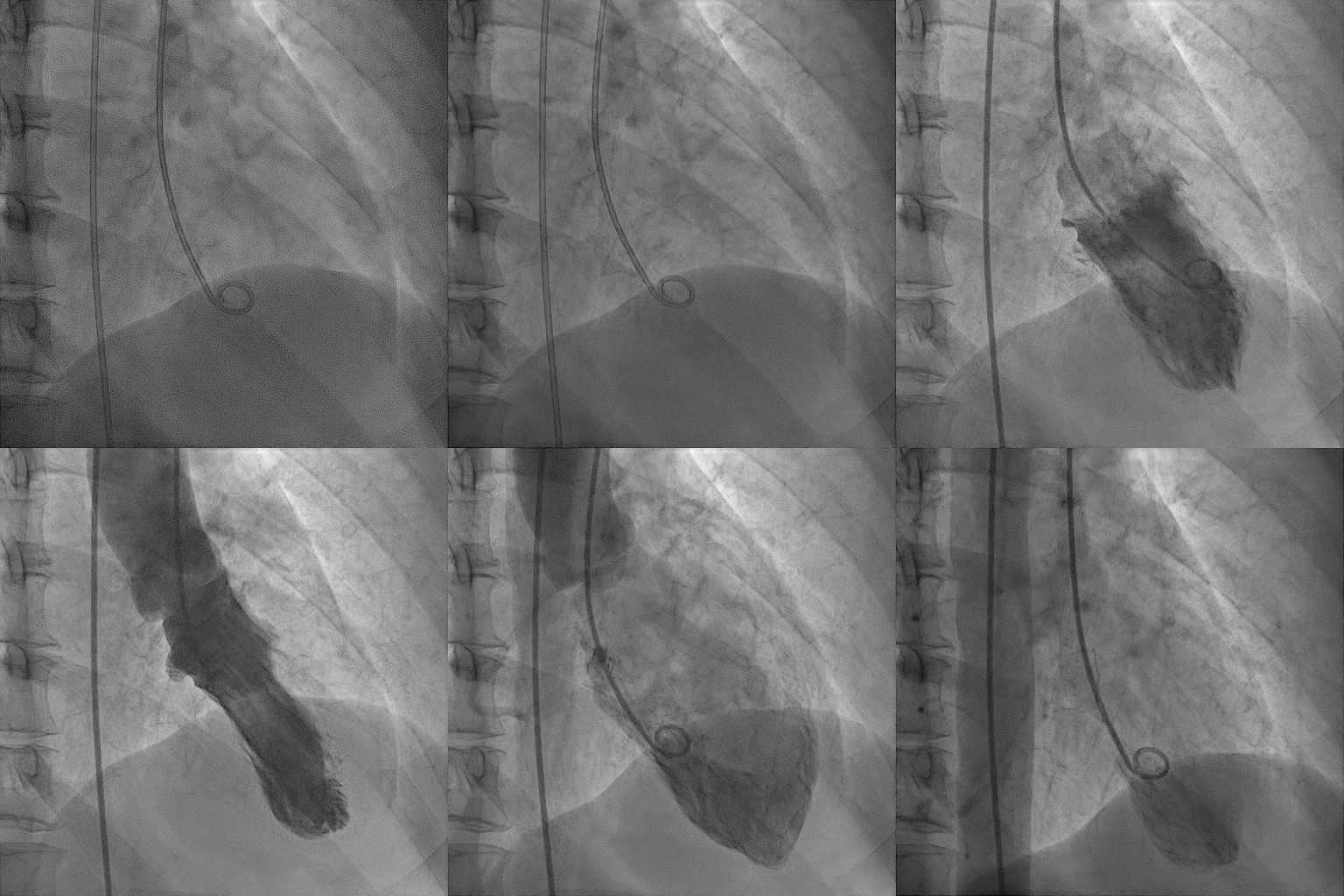
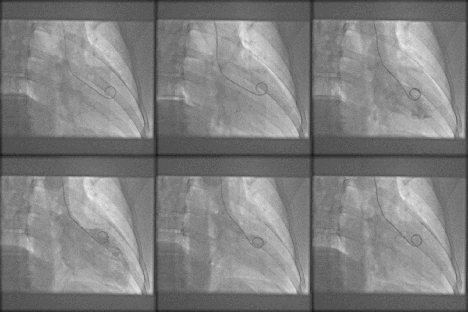
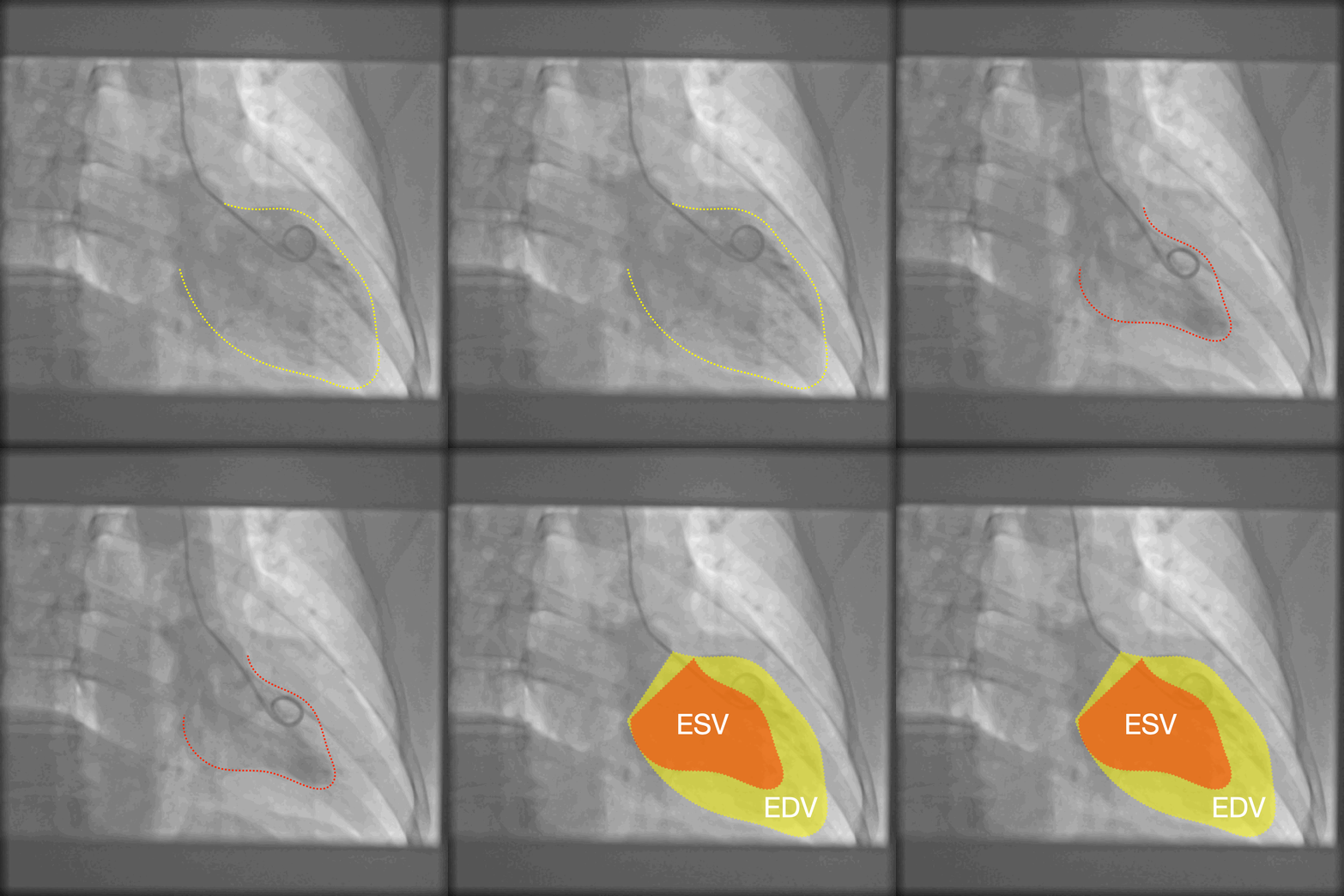
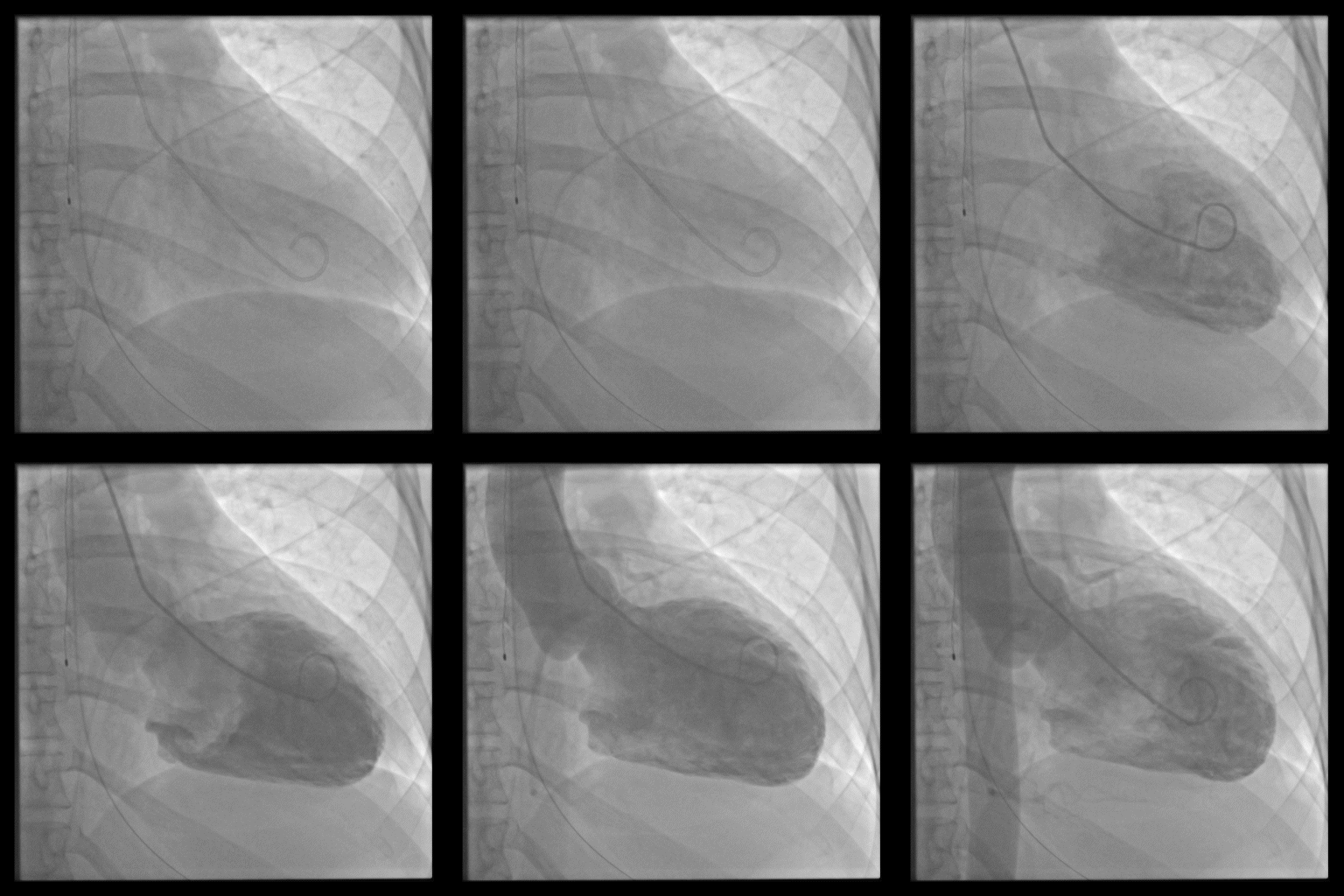
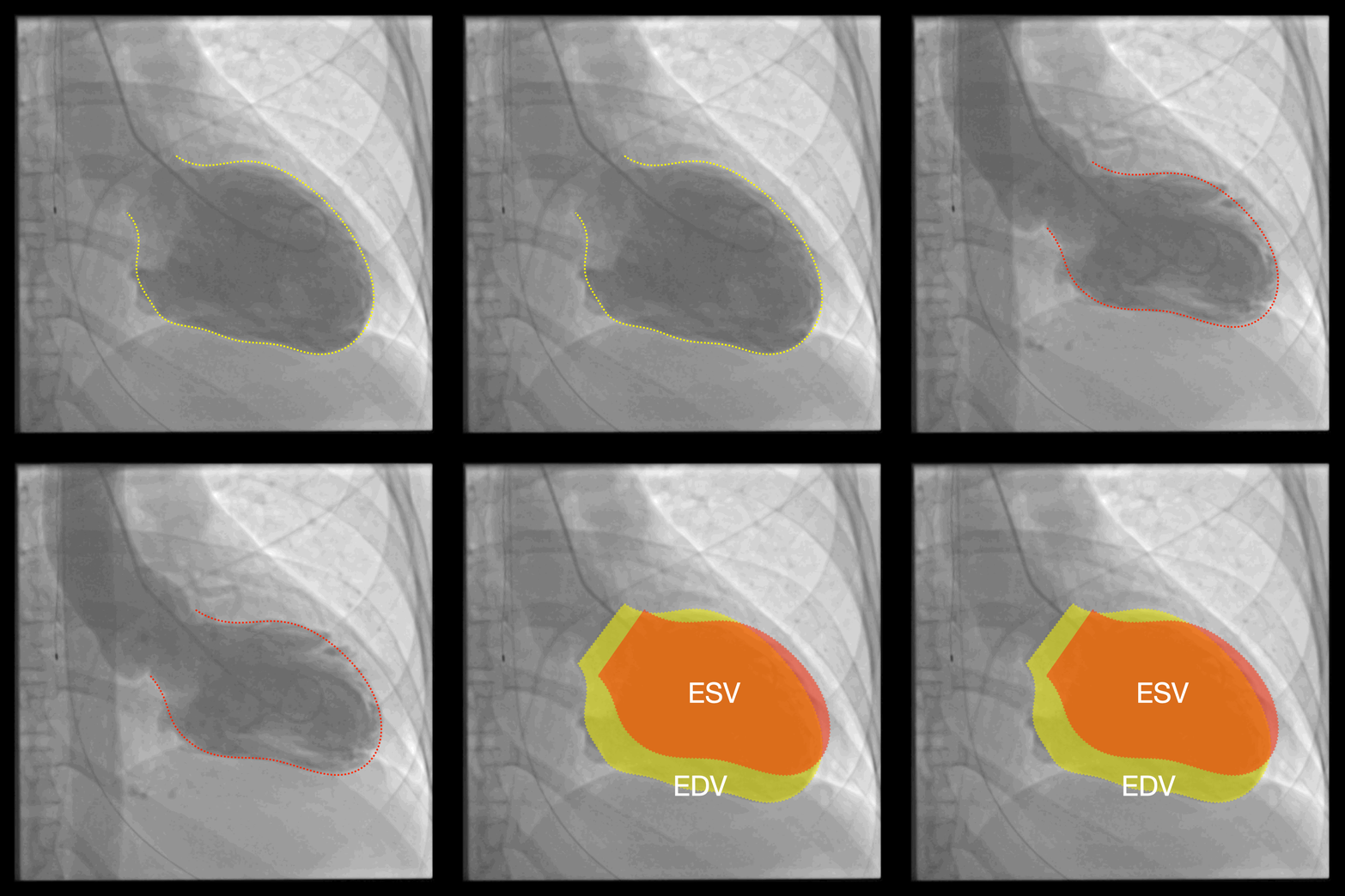
Left ventricular ejection fraction (LVEF) can be visually estimated by comparing the contour of the left ventricle (LV) between the end-systolic frame and the end-diastolic frame on left ventriculography.
Calculation
By definition, the volume of blood within a ventricle at the end of diastole is the end-diastolic volume (EDV). Likewise, the volume of blood left in a ventricle at the end of systole (contraction) is the end-systolic volume (ESV). The difference between EDV and ESV is the stroke volume (SV). The ejection fraction is the fraction of the end-diastolic volume that is ejected with each beat; that is, it is stroke volume (SV) divided by end-diastolic volume (EDV):
<math>EF (\%) = \frac{SV}{EDV}\times100</math>
Where the stroke volume is given by:
<math>SV = EDV - ESV</math>
EF is inherently a relative measurement—as is any fraction, ratio, or percentage, whereas the stroke volume, end-diastolic volume or end-systolic volume are absolute measurements.
Classification
According to the ACC Heart Failure Clinical Toolkit, based on the quantitative results of LVEF assessment, left ventricular function can be classified into the following qualitative categories:
- Hyperdynamic = LVEF greater than 70%
- Normal = LVEF 50% to 70% (midpoint 60%)
- Mild dysfunction = LVEF 40% to 49% (midpoint 45%)
- Moderate dysfunction = LVEF 30% to 39% (midpoint 35%)
- Severe dysfunction = LVEF less than 30%
Examples
Hyperdynamic (>70%)
Case 1: LVEF ≈ 75%
 |
 |
Normal LVEF (50–70%)
Case 2: LVEF ≈ 60%
 |
 |
Mild dysfunction (40–49%)
Case 3: LVEF ≈ 45%
 |
 |
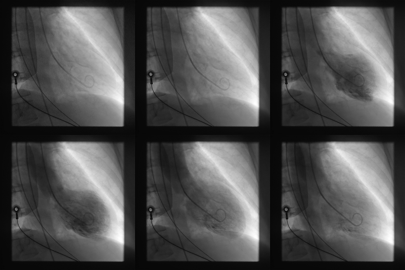
Moderate dysfunction (30–39%)
Case 4: LVEF ≈ 35%
 |
 |