Janeway lesions
| Janeway lesions | |
 | |
|---|---|
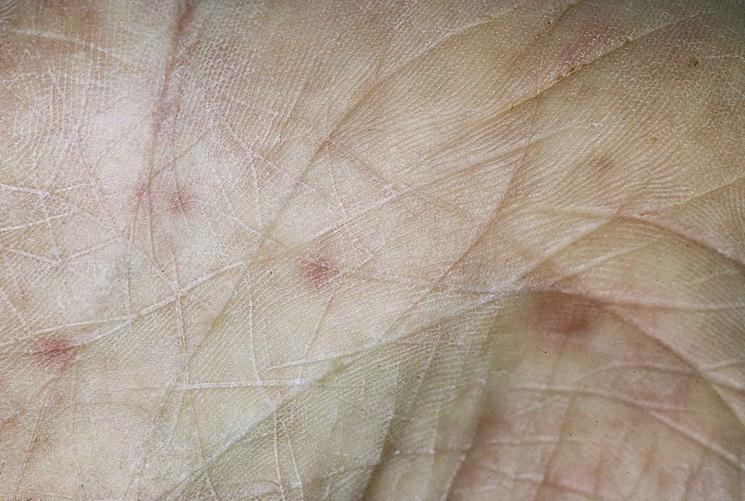
| Janeway Lesions: Flat, painless, erythematous lesions seen on the palm of this patient's hand. Frequently associated with bacterial endocarditis. (Image courtesy of Charlie Goldberg, M.D.) |
|
WikiDoc Resources for Janeway lesions |
|
Articles |
|---|
|
Most recent articles on Janeway lesions Most cited articles on Janeway lesions |
|
Media |
|
Powerpoint slides on Janeway lesions |
|
Evidence Based Medicine |
|
Clinical Trials |
|
Ongoing Trials on Janeway lesions at Clinical Trials.gov Trial results on Janeway lesions Clinical Trials on Janeway lesions at Google
|
|
Guidelines / Policies / Govt |
|
US National Guidelines Clearinghouse on Janeway lesions NICE Guidance on Janeway lesions
|
|
Books |
|
News |
|
Commentary |
|
Definitions |
|
Patient Resources / Community |
|
Patient resources on Janeway lesions Discussion groups on Janeway lesions Patient Handouts on Janeway lesions Directions to Hospitals Treating Janeway lesions Risk calculators and risk factors for Janeway lesions
|
|
Healthcare Provider Resources |
|
Causes & Risk Factors for Janeway lesions |
|
Continuing Medical Education (CME) |
|
International |
|
|
|
Business |
|
Experimental / Informatics |
Editor-In-Chief: C. Michael Gibson, M.S., M.D. [1]
Janeway lesions are non-tender, small erythematous or haemorrhagic macular or nodular lesions on the palms or soles only a few millimeters in diameter that are pathognomonic of infective endocarditis.[1] Pathologically, the lesion is described to be a microabscess of the dermis with marked necrosis and inflammatory infiltrate not involving the epidermis, which is due to the deposition of circulating immune complexes in small blood vessels.[1]
Overview
- Janeway lesions are irregular, flat, painless, erythematous macules found on the palms, soles, thenar and hypothenar eminences of the fingertips, hands and plantar surfaces of the toes.
- Stigmata of infectious endocarditis
- Considered a criterion (albeit minor) of vascular phenomena
Etymology
They are named after Edward G. Janeway (1841–1911), a professor of medicine with interests in cardiology and infectious disease.[2]
Diagnosis
(Images courtesy of Charlie Goldberg, M.D., UCSD School of Medicine and VA Medical Center, San Diego, CA)
-
 Janeway Lesions: Flat, painless, erythematous lesions seen on the palm of this patient's hand. Frequently associated with bacterial endocarditis.
 -
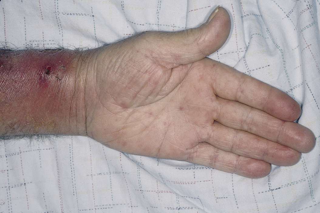 Janeway Lesion: Flat, painless, erythematous lesions seen on the palm of this patient's hand. While frequently associated with bacterial endocarditis, in this case, they are the result of an infected radial artery aneurysm (inflamed area proximal to thumb).
 -
 Janeway Lesions: Flat, painless, erythematous lesions seen on the palm of this patient's hand. Frequently associated with bacterial endocarditis.
 -
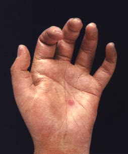 Janeway Lesions: Flat, painless, erythematous lesions seen on the palm of this patient's hand. Frequently associated with bacterial endocarditis.


History and Symptoms
- IV drug abuse
- Infective endocarditis
- Structural heart disease
- Heart valve injury/replacement
- Family history of autoimmune disorders
- HIV status
- Mucosal bleeding
- Head/neck or lung infection
- Tick exposure
- Constitutional symptoms
Laboratory Findings
- Blood cultures
- Complete blood count (CBC) with peripheral smear
- Antinuclear antibody (ANA)
- Rapid plasma reagin (RPR)
- Erythrocyte sedimentation rate (ESR)
- Antistreptolysin O antibodies (ASO)
- Urinalysis
- Coagulation studies
- Anti-SM antibodies
- Anti-dsDNA antbodies
Chest X Ray
Echocardiography or Ultrasound
Other Diagnostic Studies
- Possible biopsy
Differential Diagnosis
In alphabetical order. [3] [4]
- Acute bacterial endocarditis
- Coxsackievirus
- Cutaneous vasculitis
- Disseminated Intravascular Coagulation (DIC)
- Echovirus
- Erythema multiforme
- Idiopathic thrombocytopenia purpura
- Meningococcemia
- Polyarteritis Nodosa
- Rocky Mountain Spotted Fever
- Secondary syphilis
- Subacute bacterial endocarditis
- Systemic Lupus Erythematosus
- Thrombotic thrombocytopenic purpura
- Typhoid Fever
Acute Pharmacotherapies
- IV antibiotics
- Antibiotic prophylaxis
- Treat all underlying etiologies
Chronic Pharmacotherapies
- Antibiotic therapy for bacterial endocarditis
Surgery and Device Based Therapy
- Valve replacement for bacterial endocarditis
Indications for Surgery
- If all other medical therapy fails (bacterial endocarditis)
References
- ↑ 1.0 1.1 Farrior, J.B. (1976). "A consideration of the differences between a Janeway's lesion and an Osler's node in infectious endocarditis". Chest. 70 (2): 239–43. doi:10.1378/chest.70.2.239. PMID 947688. Unknown parameter
|coauthors=ignored (help) - ↑ Janeway C. (1998). "Presidential Address to The American Association of Immunologists. The road less traveled by: the role of innate immunity in the adaptive immune response". J. Immunol. 161 (2): 539–44. PMID 9670925.
- ↑ Sailer, Christian, Wasner, Susanne. Differential Diagnosis Pocket. Hermosa Beach, CA: Borm Bruckmeir Publishing LLC, 2002:77 ISBN 1591032016
- ↑ Kahan, Scott, Smith, Ellen G. In A Page: Signs and Symptoms. Malden, Massachusetts: Blackwell Publishing, 2004:68 ISBN 140510368X
See also
Template:Eponymous medical signs for circulatory and respiratory systems