Sandbox:DAMI
RESIDENT SURVIVAL GUIDE
| Resident Survival Guide |
|---|
| Introduction |
| Team |
| Guide |
| Page Template |
| Examine the Patient Template |
| Navigation Bar Template |
| Checklist |
| Topics |


Editor-In-Chief: C. Michael Gibson, M.S., M.D. [1]; Associate Editor(s)-in-Chief: Mahmoud Sakr, M.D. [2]
Differential diagnosis of abdominal pain
Abbreviations: RUQ= Right upper quadrant of the abdomen, LUQ= Left upper quadrant, LLQ= Left lower quadrant, RLQ= Right lower quadrant, LFT= Liver function test, SIRS= Systemic inflammatory response syndrome, ERCP= Endoscopic retrograde cholangiopancreatography, IV= Intravenous, N= Normal, AMA= Anti mitochondrial antibodies, LDH= Lactate dehydrogenase, GI= Gastrointestinal, CXR= Chest X ray, IgA= Immunoglobulin A, IgG= Immunoglobulin G, IgM= Immunoglobulin M, CT= Computed tomography, PMN= Polymorphonuclear cells, ESR= Erythrocyte sedimentation rate, CRP= C-reactive protein, TS= Transferrin saturation, SF= Serum Ferritin, SMA= Superior mesenteric artery, SMV= Superior mesenteric vein, ECG= Electrocardiogram
| |||||||||||||||||||||||||||||||||||||||||||||||||||||||||||||||||||||||||||||||||||||||||||||||||||||||||||||||||||||||||||||||||||||||||||||||||||||||||||||||||||||||||||||||||||||||||||||||||||||||||||||||||||||||||||||||||||||||||||||||||||||||||||||||||||||||||||||||||||||||||||||||||||||||||||||||||||||||||||||||||||||||||||||||||||||||||||||||||||||||||||||||||||||||||||||||||||||||||||||||||||||||||||||||||||||||||||||||||||||||||||||||||||||||||||||||||||||||||||||||||||||||||||||||||||||||||||||||||||||||||||||||||||||||||||||||||||||||||||||||||||||||||||||||||||||||||||||||||||||||||||||||||||||||||||||||||||||||||||||||||||||||||||||||||||||||||||||||||||||||||||||||||||||||||||||||||||||||||||||||||||||||||||||||||||||||||||||||||||||||||||||||||||||||||||||||||||||||||||||||||||||||||||||||||||||||||||||||||||||||||||||||||||||||||||||||||||||||||||||||||||||||||||||||||||||||||||||||||||||||||||||||||||||||||||||||||||||||||||||||||||||||||||||||||||||||||||||||||||||||
<figure-inline class="mw-default-size"><figure-inline> </figure-inline></figure-inline> </figure-inline></figure-inline> |
<figure-inline class="mw-default-size"><figure-inline> </figure-inline></figure-inline> </figure-inline></figure-inline> |
<figure-inline class="mw-default-size"><figure-inline>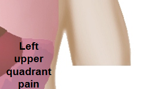 </figure-inline></figure-inline> </figure-inline></figure-inline>
|
<figure-inline class="mw-default-size"><figure-inline> </figure-inline></figure-inline> </figure-inline></figure-inline> |
<figure-inline class="mw-default-size"><figure-inline> </figure-inline></figure-inline> </figure-inline></figure-inline> |
<figure-inline class="mw-default-size"><figure-inline> </figure-inline></figure-inline> </figure-inline></figure-inline>
|
<figure-inline class="mw-default-size"><figure-inline>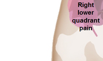 </figure-inline></figure-inline> </figure-inline></figure-inline> |
<figure-inline class="mw-default-size"><figure-inline>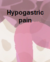 </figure-inline></figure-inline> </figure-inline></figure-inline> |
<figure-inline class="mw-default-size"><figure-inline>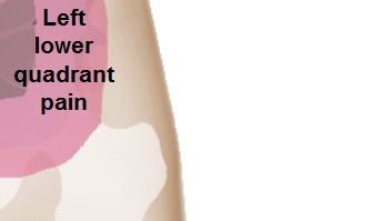 </figure-inline></figure-inline> </figure-inline></figure-inline>
|
The following is a list of diseases that present with acute onset severe lower abdominal pain:
| Disease | Findings |
|---|---|
| Ectopic pregnancy | History of missed menses, positive pregnancy test, ultrasound reveals an empty uterus and may show a mass in the fallopian tubes.[1] |
| Appendicitis | Pain localized to the right iliac fossa, vomiting, abdominal ultrasound sensitivity for diagnosis of acute appendicitis is 75% to 90%.[2] |
| Rupturedovarian cyst | Usually spontaneous, can follow history of trauma, mild chronic lower abdominal discomfort may suddenly intensify, ultrasound is diagnostic.[3] |
| Ovarian cyst torsion | Presents with acute severe unilateral lower quadrant abdominal pain, nausea and vomiting, tender adnexal mass palpated in 90%, ultrasound is diagnostic.[4] |
| Hemorrhagic ovarian cyst | Presents with localized abdominal pain, nausea and vomiting. Hypovolemic shock may be present, abdominal tenderness and guarding are physical exam findings, ultrasound is diagnostic.[4] |
| Endometriosis | Presents with cyclic pain that is exacerbated by onset of menses, dyspareunia. laparoscopic exploration is diagnostic.[4] |
| Acute cystitis | Presents with features of increased urinary frequency, urgency, dysuria, and suprapubic pain.[5][6] |
References
- ↑ Morin L, Cargill YM, Glanc P (2016). "Ultrasound Evaluation of First Trimester Complications of Pregnancy". J Obstet Gynaecol Can. 38 (10): 982–988. doi:10.1016/j.jogc.2016.06.001. PMID 27720100.
- ↑ Balthazar EJ, Birnbaum BA, Yee J, Megibow AJ, Roshkow J, Gray C (1994). "Acute appendicitis: CT and US correlation in 100 patients". Radiology. 190 (1): 31–5. doi:10.1148/radiology.190.1.8259423. PMID 8259423.
- ↑ Bottomley C, Bourne T (2009). "Diagnosis and management of ovarian cyst accidents". Best Pract Res Clin Obstet Gynaecol. 23 (5): 711–24. doi:10.1016/j.bpobgyn.2009.02.001. PMID 19299205.
- ↑ 4.0 4.1 4.2 Bhavsar AK, Gelner EJ, Shorma T (2016). "Common Questions About the Evaluation of Acute Pelvic Pain". Am Fam Physician. 93 (1): 41–8. PMID 26760839.
- ↑ {{Cite journal | author = W. E. Stamm | title = Etiology and management of the acute urethral syndrome | journal = Sexually transmitted diseases | volume = 8 | issue = 3 | pages = 235–238 | year = 1981 | month = July-September | pmid = 7292216
- ↑ {{Cite journal | author = W. E. Stamm, K. F. Wagner, R. Amsel, E. R. Alexander, M. Turck, G. W. Counts & K. K. Holmes | title = Causes of the acute urethral syndrome in women | journal = The New England journal of medicine | volume = 303 | issue = 8 | pages = 409–415 | year = 1980 | month = August | doi = 10.1056/NEJM198008213030801 | pmid = 6993946
Causes
Life Threatening Causes
- Central Diabetes Insipidus
- Nephrogenic Diabetes insipidus
- Psychogenic Diabetes insipidus
- Gestational Diabetes insipidus
- Autoimmune Diabetes insipidus
Common Causes
Diagnosis
Shown below is an algorithm summarizing the diagnosis of [[disease name]] according the the [...] guidelines.
Treatment
Shown below is an algorithm summarizing the treatment of [[disease name]] according the the [...] guidelines.
Do's
- The content in this section is in bullet points.
Don'ts
- The content in this section is in bullet points.
References
Differentiating Protein energy malnutrition from other Diseases
Differentiating kwashiorkor from marasmus[1]
| Distinguishing Features | Kwashiorkor | Marasmus |
|---|---|---|
| Cause | Deficiency of protein in the diet of child | Deficiency of protein as well as energy nutrients (that is carbohydrates and fats) in the diet |
| Age | Occurs in children in the age group 1-5 years | Typically occurs in children below the age of 1 year |
| Association | More common in villages where there is small gap period between successive pregnancies | More common in towns and cities where breast-feeding in discontinued quite early |
| Edema | Presence of edema | Absence of edema |
| Muscles | Wasting of muscles | Wasting of muscles is quite evident. The child is reduced to skin and bones |
| Skin changes | Dermatitis and Hyperpigmentation noticed | Dry and atrophic skin but no changes in color |
| Serum cortisol | Decreased/Normal | Increased |
| Fasting blood glucose | Decreased | Decreased |
| Growth retardation | Mildly retarded in growth | Severely retarded in growth |
| Facial appearance | Moon-like face | Elderly man face |
| Abdomen | Protuded | Shrunken |
| Vitamin deficiency | Present | Present |
| Weight | 60-80% of normal weight for age | <60% of normal weight for age |
Differential diagnosis of childhood malnutrition
| Kwashiorkor | |||
| Marasmus | |||
| Vitamin A deficiency | |||
| Iron deficiency | |||
| Iodine deficiency | |||
| Congenital heart disease | |||
| Dehydration | |||
| Sepsis | |||
| Shigella and campylobacter | |||
| Inflammatory bowel diseases |
Causes in Alphabetical Order
|
|
|
Causes by Organ System
| Cardiovascular | Hypovolemic shock |
| Chemical / poisoning | No underlying causes |
| Dermatologic | No underlying causes |
| Drug Side Effect | Amphotericin B, Demeclocycline, Lithium , Radiation - for CNS and nasopharyngeal malignancies , Pituitary radiation , Netilmicin , Kanamycin, Carbamazepine , Amikacin |
| Ear Nose Throat | No underlying causes |
| Endocrine | Ischemia of the pitutary gland , Sheehan syndrome , Pituitary tumour , Panhypopituitarism , lymphocytic hypophysitis , Familial hypopituitarism , congenital nephrogenic , Congenital hypopituitarism , Apoplexy , pituitary cysts |
| Environmental | No underlying causes |
| Gastroenterologic | No underlying causes |
| Genetic | Inherited genetic kidney disorders , Genetic diseases - pit-1 mutation |
| Hematologic | Sickle cell disease , Langerhans cell histiocytosis |
| Iatrogenic | Neurosurgery , Pituitary surgery |
| Infectious Disease | Tuberculous meningitis , Meningoencephalitis , Infections |
| Musculoskeletal / Ortho | No underlying causes |
| Neurologic | Dipsogenic diabetes insipidus , Ischemic encephalopathy , Subarachnoid hemorrhage , Mass lesions hypothalamus , Ischemic stroke , Intracranial space-occupying lesion , Intracranial bleeding , Hypoxic encephalopathy , Hypothalamic radiation , Brain aneurysm |
| Nutritional / Metabolic | Hypercalcemia , hemochromatosis , Anorexia nervosa |
| Obstetric/Gynecologic | Gestational diabetes insipidus |
| Oncologic | Pituitary Cancer , pituitary adenomas , malignant tumors infilterating hypothalamus from lung and breast , Histiocytosis , Craniopharyngioma |
| Opthalmologic | No underlying causes |
| Overdose / Toxicity | No underlying causes |
| Psychiatric | No underlying causes |
| Pulmonary | No underlying causes |
| Renal / Electrolyte | Polycystic kidney disease, Renal failure , Interstitial nephritis , Hypokalaemia , Hypercalcaemia |
| Rheum / Immune / Allergy | Sarcoidosis , Autoimmune Hypophysitis , Autoimmune Diabetes Insipidus |
| Sexual | No underlying causes |
| Trauma | Head injury , Trauma - fracture of skull base |
| Urologic | Partial blockage of the ureters |
| Dental | No underlying causes |
| Miscellaneous | Dehydration , Wolfram syndrome , Sicca syndrome , Septo-optic dysplasia , Infiltrative lesions , Idiopathic , Hand-Schuller-Christian Syndrome , Familial CDI , Erdheim-Chester disease , DIDMOAD syndrome , Anophthalmia -- hypyothalamo-pituitary insufficiency , Anophthalmia -- hypothalamo-pituitary insufficiency , Amyloidosis |
Table for HV
- The table below summarizes the classification of the herpesviridae family
| Classification | Clinical manifestations | |
|---|---|---|
| Subfamily
alphavirinae |
Herpes simplex type 1 |
|
| Herpes simplex type 2
varicella-zoster virus |
| |
| Subfamily betavirinae | Cytomegalovirus |
Fever
|
| Human herpes virus 6 | Acute febrile illness sometimes with rash (roseola infantum) | |
| Human herpes virus 7 | May cause febrile illness sometimes with rash ( roseola-like) | |
| Subfamily
gammavirinae |
Epstein-Bar virus | mononucleosis, lymphoma, nasopahryngeal carcinoma and hodgkins disease. |
| Human herpes virus 8 | Kaposi's sarcoma in immunocompromised. |
Watery diarrhea
- Osmotic diarrhea
- Mg2+, PO43-, SO42- ingestion
- Carbohydrate malabsorption
- Secretory diarrhea
- Laxative abuse (nonosmotic laxatives)
- Congenital syndromes
- Bacterial toxins
- Ileal bile acid malabsorption
- Inflammatory bowel disease
- Ulcerative colitis
- Crohn’s disease
- Microscopic (lymphocytic and collagenous) colitis
- Diverticulitis
- Vasculitis
- Drugs and poisons
- Disordered motility
- Postvagotomy diarrhea
- Postsympathectomy diarrhea
- Diabetic autonomic neuropathy
- Hyperthyroidism
- Irritable bowel syndrome
- Neuroendocrine tumors
- Gastrinoma
- VIPoma
- Somatostatinoma
- Mastocytosis
- Carcinoid syndrome
- Medullary carcinoma of thyroid
- Neoplasia
- Colon carcinoma
- Lymphoma
- Villous adenoma
- Addison’s disease
- Epidemic secretory diarrhea
- Idiopathic secretory diarrhea
Fatty diarrhea
- Malabsorption syndromes
- Mucosal diseases
- Short-bowel syndrome
- Postresection diarrhea
- Mesenteric ischemia
- Maldigestion
- Pancreatic insufficiency
- Bile acid deficiency
Inflammatory diarrhea
- Inflammatory bowel disease
- Ulcerative colitis
- Crohn’s disease
- Diverticulitis
- Ulcerative jejunoileitis
- Infectious diseases
- Ulcerating viral infections
- Cytomegalovirus
- Herpes simplex
- Ulcerating viral infections
- Ischemic colitis
- Radiation colitis
- Neoplasia
- Colon cancer
- Lymphoma
| Cause | Osmotic gap | History | Physical exam | Labs | Gold standard | Treatment | |||
|---|---|---|---|---|---|---|---|---|---|
| Osmotic gap | Other Labs | ||||||||
| Watery | Secretory | Crohns | |||||||
| IBS | |||||||||
| Osmotic | |||||||||
| FunctionL | |||||||||
Watery
- Secretory (often nocturnal; unrelated to food intake; fecal osmotic gap < 50 mOsm per kg*)
- Alcoholism
- Bacterial enterotoxins (e.g., cholera)
- Bile acid malabsorption
- Brainerd diarrhea (epidemic secretory diarrhea)
- Congenital syndromes
- Crohn disease (early ileocolitis)
- Endocrine disorders e.g., hyperthyroidism
- Medications (see causes section)
- Microscopic colitis (lymphocytic and collagenous subtypes)
- Neuroendocrine tumors (e.g., gastrinoma, vipoma, carcinoid tumors, mastocytosis)
- Nonosmotic laxatives (e.g., senna, docusate sodium)
- Postsurgical (e.g., cholecystectomy, gastrectomy, vagotomy, intestinal resection)
- Vasculitis
- Osmotic (fecal osmotic gap > 50 mOsm per kg*)
- Carbohydrate malabsorption syndromes (e.g., lactose, fructose)
- Celiac disease
- Osmotic laxatives and antacids (e.g., magnesium, phosphate, sulfate)
- Sugar alcohols (e.g., mannitol, sorbitol, xylitol)
- Functional (distinguished from secretory types by hypermotility, smaller volumes, and improvement at night and with fasting)
Table showing watery causes of chronic diarrhea (Table 1)
| Cause | Osmotic gap | History | Physical exam | Gold standard | Treatment | |||
|---|---|---|---|---|---|---|---|---|
| < 50 mOsm per kg | > 50 mOsm per kg* | |||||||
| Watery | Secretory | Crohns | + | - |
|
|
|
|
| Hyperthyroidism | + | - |
|
|
||||
| VIPoma | + | - |
|
|
|
| ||
| Osmotic | Lactose intolerance | - | + |
|
||||
| Celiac disease | - | + |
|
|
|
|||
| Functional | Irritable bowel syndrome | - | - |
Abdominal pain or discomfort recurring at least 3 days per month in the past 3 months and associated with 2 or more of the following:
History of straining is also common |
|
|
| |
Fatty (bloating and steatorrhea in many, but not all cases)
- Malabsorption syndrome (damage to or loss of absorptive ability)
- Amyloidosis
- Carbohydrate malabsorption (e.g., lactose intolerance)
- Celiac sprue (gluten enteropathy)–various clinical presentations
- Gastric bypass
- Lymphatic damage (e.g., congestive heart failure, some lymphomas)
- Medications (e.g., orlistat Xenical; inhibits fat absorption, acarbose Precose; inhibits carbohydrate absorption])
- Mesenteric ischemia
- Noninvasive small bowel parasite (e.g., Giardia)
- Post-resection diarrhea
- Short bowel syndrome
- Small bowel bacterial overgrowth (> 105 bacteria per mL)
- Tropical sprue
- Whipple disease (Tropheryma whippelii infection)
- Maldigestion (loss of digestive function)
- Hepato-biliary disorders
- Inadequate luminal bile acid
- Loss of regulated gastric emptying
- Pancreatic exocrine insufficiency
Table showing fatty causes of chronic diarrhea ( Table 2)
| Cause | Osmotic gap | History | Physical exam | Gold standard | Treatment | |
|---|---|---|---|---|---|---|
| < 50
mOsm per kg |
> 50
mOsm per kg* | |||||
| lactose intolerance | - | + |
|
|
Lactose breath hydrogen test | Restriction of lactose and maintain calcium and vitamin D intake. |
| Celiac sprue | - | + |
|
|
Immunoglobulin A (IgA) anti-tissue transglutaminase (TTG) antibody followed by upper ebdoscopy with biopsy. | Dietary counseling, elimination of gluten in the diet. |
| Whipple disease | - | + |
|
|
Upper endoscopy with biopsies of the small intestine for T. whipplei testing (histology with PAS staining, polymerase chain reaction [PCR] testing, and immunohistochemistry) | Doxycycline and hydroxychloroquine was bactericidal |
Inflammatory or exudative (elevated white blood cell count, occult or frank blood or pus)
- Inflammatory bowel disease Crohn disease (ileal or early Crohn disease may be secretory)
- Diverticulitis
- Ulcerative colitis
- Ulcerative jejunoileitis
- Invasive infectious diseases
- Clostridium difficile (pseudomembranous) colitis–antibiotic history
- Invasive bacterial infections (e.g., tuberculosis, yersiniosis)
- Invasive parasitic infections (e.g., Entamoeba)–travel history
- Ulcerating viral infections (e.g., cytomegalovirus, herpes simplex virus)
- Neoplasia
- Colon carcinoma
- Lymphoma
- Villous adenocarcinoma
- Radiation colitis
Table showing inflammatory causes of chronic diarrhea ( Table 3)
| Cause | History | Laboratory findings | Diagnosis | Treatment |
|---|---|---|---|---|
| Diverticulitis |
|
|
Abdominal CT scan with oral and intravenous (IV) contrast | bowel rest, IV fluid resuscitation, and broad-spectrum antimicrobial therapy which covers anaerobic bacteria and gram-negative rods |
| Ulcerative colitis |
|
|
Endoscopy | Induction of remission with mesalamine and corticosteroids followed by the administration of sulfasalazine and 6-Mercaptopurine depending on the severity of the disease. See ... |
| Entamoeba histolytica |
|
cysts shed with the stool | detects ameba DNA in feces | Amebic dysentery ;
Luminal amebicides for E. histolytica in the colon:
For amebic liver abscess:
|
==Gestational Diabetes insipidus
Differentiating Diabetes insipidus based on the levels of ADH and the response of the body to the level of hyponatremia
- Disorders in which ADH levels are elevated[2]
- Reduced effective arterial blood volume
- True volume depletion
- Heart failure
- Cirrhosis
- Syndrome of inappropriate ADH secretion, including reset osmostat pattern
- Hormonal changes
- Reduced effective arterial blood volume
- Disorders in which ADH levels may be appropriately suppressed[3]
- Advanced renal failure
- Primary polydipsia
- Diabetes mellitus
- Sickle cell disease
- Hyponatremia with normal or elevated plasma osmolality[4]
- High plasma osmolality (effective osmols)
- High plasma osmolality (ineffective osmols)
- Renal failure
- Alcohol intoxication with an elevated serum alcohol concentration
- Normal plasma osmolality
- Pseudohyponatremia (laboratory artifact)
- Absorption of irrigant solutions
Question on Roseola
- A woman brings her 14 month old baby to the physician for the evaluation of a rash. He was in a good state of health until about 3 days ago when he developed a very high fever. The mother says the temperature was as high as 40C (104F) when she measured it with her thermometer at home. She gave him some tylenol and the fever subsided after which the rash developed. It started as a non itchy pink rash with rose spots on the head and is now generalized all over the body. Today the boy's temperature measured in the clinic is 37 C( 98F), pulse 88/min and respirations are 16/min. His immunizations are up to date and the boy is in no apparent distress. What is the most likely diagnosis in this patient?
- A. Scarlet Fever
- B. Rubella(German measles
- C. Roseola (sixth disease)
- D. Rocky mountain spotted fever
- E. Measles
- F. Kawasaki disease
- G. Erythema infectiosum (fifth disease)
- ↑ Müller O, Krawinkel M (2005). "Malnutrition and health in developing countries". CMAJ. 173 (3): 279–86. doi:10.1503/cmaj.050342. PMC 1180662. PMID 16076825.
- ↑ Danziger J, Zeidel ML (2015). "Osmotic homeostasis". Clin J Am Soc Nephrol. 10 (5): 852–62. doi:10.2215/CJN.10741013. PMC 4422250. PMID 25078421.
- ↑ Sterns RH (2015). "Disorders of plasma sodium--causes, consequences, and correction". N Engl J Med. 372 (1): 55–65. doi:10.1056/NEJMra1404489. PMID 25551526.
- ↑ Fenske WK, Christ-Crain M, Hörning A, Simet J, Szinnai G, Fassnacht M; et al. (2014). "A copeptin-based classification of the osmoregulatory defects in the syndrome of inappropriate antidiuresis". J Am Soc Nephrol. 25 (10): 2376–83. doi:10.1681/ASN.2013080895. PMC 4178436. PMID 24722436.