Gram-negative
|
WikiDoc Resources for Gram-negative |
|
Articles |
|---|
|
Most recent articles on Gram-negative Most cited articles on Gram-negative |
|
Media |
|
Powerpoint slides on Gram-negative |
|
Evidence Based Medicine |
|
Clinical Trials |
|
Ongoing Trials on Gram-negative at Clinical Trials.gov Trial results on Gram-negative Clinical Trials on Gram-negative at Google
|
|
Guidelines / Policies / Govt |
|
US National Guidelines Clearinghouse on Gram-negative NICE Guidance on Gram-negative
|
|
Books |
|
News |
|
Commentary |
|
Definitions |
|
Patient Resources / Community |
|
Patient resources on Gram-negative Discussion groups on Gram-negative Patient Handouts on Gram-negative Directions to Hospitals Treating Gram-negative Risk calculators and risk factors for Gram-negative
|
|
Healthcare Provider Resources |
|
Causes & Risk Factors for Gram-negative |
|
Continuing Medical Education (CME) |
|
International |
|
|
|
Business |
|
Experimental / Informatics |
Editor-In-Chief: C. Michael Gibson, M.S., M.D. [1]
Overview

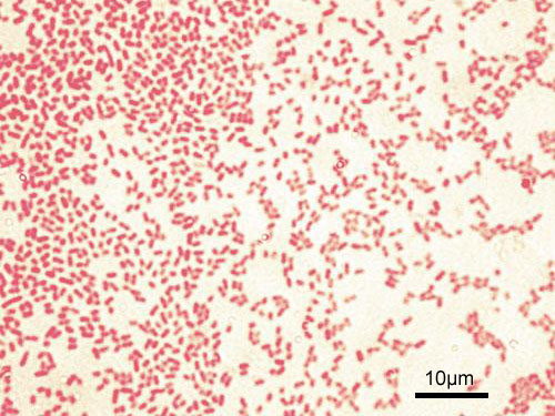
Gram-negative bacteria are those that do not retain crystal violet dye in the Gram staining protocol.[1] Gram-positive bacteria will retain the dark blue dye after an alcohol wash. In a Gram stain test, a counterstain (commonly Safranin) is added after the crystal violet, colouring all Gram-negative bacteria a red or pink colour. The test itself is useful in classifying two distinctly different types of bacteria based on structural differences in their cell walls.[2]
Many species of Gram-negative bacteria are pathogenic, meaning they can cause disease in a host organism. This pathogenic capability is usually associated with certain components of Gram-negative cell walls, in particular the lipopolysaccharide (also known as LPS or endotoxin) layer.[1] The LPS is the trigger which the body's innate immune response receptors sense to begin a cytokine reaction. It is toxic to the host. It is this response which begins the inflammation cycle in tissues and blood vessels.
Characteristics

The following characteristics are displayed by Gram-negative bacteria:
- Cell walls only contain a few layers of peptidoglycan (which is present in much higher levels in Gram-positive bacteria)
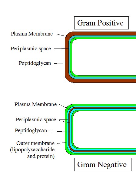
- Cells are surrounded by an outer membrane containing lipopolysaccharide (which consists of Lipid A, core polysaccharide, and O-polysaccharide) outside the peptidoglycan layer
- Porins exist in the outer membrane, which act like pores for particular molecules
- There is a space between the layers of peptidoglycan and the secondary cell membrane called the periplasmic space
- The S-layer is directly attached to the outer membrane, rather than the peptidoglycan
- If present, flagella have four supporting rings instead of two
- No teichoic acids or lipoteichoic acids are present
- Lipoproteins are attached to the polysaccharide backbone whereas in Gram-positive bacteria no lipoproteins are present
- Most do not sporulate (Coxiella burnetti forms spore-like structures).
Example species
The proteobacteria are a major group of Gram-negative bacteria, including Escherichia coli, Salmonella, and other Enterobacteriaceae, Pseudomonas, Moraxella, Helicobacter, Stenotrophomonas, Bdellovibrio, acetic acid bacteria, Legionella and alpha-proteobacteria as Wolbachia and many others. Other notable groups of Gram-negative bacteria include the cyanobacteria, spirochaetes, green sulfur and green non-sulfur bacteria. Crenarchaeota: Unique because most bacteria have gram-positive molecules in their capsules, it has gram-negative.
Medically relevant Gram-negative cocci include three organisms, which cause a sexually transmitted disease (Neisseria gonorrhoeae), a meningitis (Neisseria meningitidis), and respiratory symptoms (Moraxella catarrhalis).
Medically relevant Gram-negative bacilli include a multitude of species. Some of them primarily cause respiratory problems (Hemophilus influenzae, Klebsiella pneumoniae, Legionella pneumophila, Pseudomonas aeruginosa), primarily urinary problems (Escherichia coli, Proteus mirabilis, Enterobacter cloacae, Serratia marcescens), and primarily gastrointestinal problems (Helicobacter pylori, Salmonella enteritidis, Salmonella typhi).
Nosocomial gram negative bacteria include Acinetobacter baumanii, which cause bacteremia, secondary meningitis, and ventilator-associated pneumonia in intensive care units of hospital establishments.
Medical treatment
One of the several unique characteristics of Gram-negative bacteria is the outer membrane. This outer membrane is responsible for protecting the bacteria from several antibiotics, dyes, and detergents which would normally damage the inner membrane or cell wall (peptidoglycan). The outer membrane provides these bacteria with resistance to lysozyme and penicillin. Fortunately, alternative medicinal treatments such as lysozyme with EDTA, and the antibiotic ampicillin have been developed to combat the protective outer membrane of some pathogenic Gram-negative organisms. Other drugs can be used, namely chloramphenicol, streptomycin, and nalidixic acid.
See also
- Gram-positive (Microbiology)
- Braun's lipoprotein (Microbiology)
References
- ↑ 1.0 1.1 Salton MJR, Kim KS (1996). Structure. in: Baron's Medical Microbiology (Baron S et al, eds.) (4th ed. ed.). Univ of Texas Medical Branch. ISBN 0-9631172-1-1.
- ↑ Madigan M, Martinko J (editors) (2005). Brock Biology of Microorganisms (11th ed. ed.). Prentice Hall. ISBN 0-13-144329-1.
External links
br:Gram-nann cs:Gramnegativní bakterie id:Gram-negatif it:Gram - nl:Gram-negatieve bacteriën no:Gram-negative sl:Gramnegativne bakterije fi:Gramnegatiivinen bakteeri uk:Грам-негативні бактерії