Gram-positive
|
WikiDoc Resources for Gram-positive |
|
Articles |
|---|
|
Most recent articles on Gram-positive Most cited articles on Gram-positive |
|
Media |
|
Powerpoint slides on Gram-positive |
|
Evidence Based Medicine |
|
Clinical Trials |
|
Ongoing Trials on Gram-positive at Clinical Trials.gov Trial results on Gram-positive Clinical Trials on Gram-positive at Google
|
|
Guidelines / Policies / Govt |
|
US National Guidelines Clearinghouse on Gram-positive NICE Guidance on Gram-positive
|
|
Books |
|
News |
|
Commentary |
|
Definitions |
|
Patient Resources / Community |
|
Patient resources on Gram-positive Discussion groups on Gram-positive Patient Handouts on Gram-positive Directions to Hospitals Treating Gram-positive Risk calculators and risk factors for Gram-positive
|
|
Healthcare Provider Resources |
|
Causes & Risk Factors for Gram-positive |
|
Continuing Medical Education (CME) |
|
International |
|
|
|
Business |
|
Experimental / Informatics |
Editor-In-Chief: C. Michael Gibson, M.S., M.D. [1]
Overview

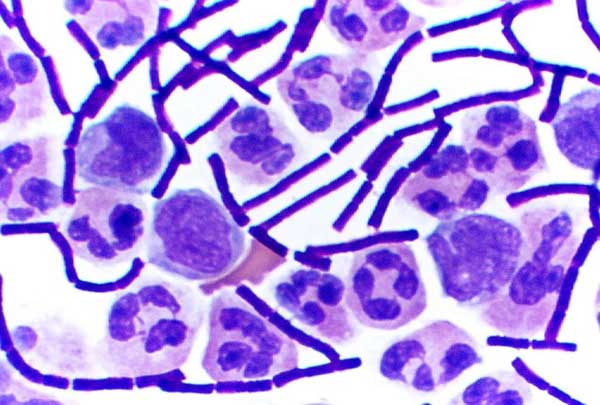
Gram-positive bacteria are those that retain a crystal violet dye during the Gram stain process.[1] Gram-positive bacteria appear blue or violet under a microscope, while Gram-negative bacteria appear red or pink. The Gram classification system is empirical, and largely based on differences in cell wall structure.[2] The purpose of Gram staining is to visually differentiate groups of bacteria, primarily for identification.
Characteristics

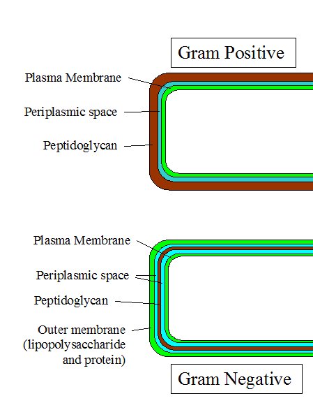
The following characteristics are generally present in a Gram-positive bacterium:[3]
- A very thick cell wall (peptidoglycan).
- If a flagellum is present, it contains two rings for support as opposed to four in Gram-negative bacteria because Gram-positive bacteria have only one membrane layer.
- Teichoic acids and lipoteichoic acids are present, which serve to act as chelating agents, and also for certain types of adherence.

History of Gram positive
In the original bacterial phyla, the Gram-positive forms made up the phylum Firmicutes, a name now used for the largest group. It includes many well-known genera such as Bacillus, Listeria, Staphylococcus, Streptococcus, Enterococcus, and Clostridium. It has also been expanded to include the Mollicutes, bacteria like Mycoplasma that lack cell walls and so cannot be stained by Gram, but are derived from such forms.
The actinobacteria are another major group of Gram-positive bacteria; they and the Firmicutes are referred to as the high and low G+C groups based on the guanosine and cytosine content of their DNA. If the second membrane is a derived condition, the two may have been basal among the bacteria; otherwise they are probably a relatively recent monophyletic group. They have been considered as possible ancestors for the archaeans and eukaryotes, both because they are unusual in lacking the second membrane and because of various biochemical similarities such as the presence of sterols.
The Deinococcus-Thermus bacteria also have Gram-positive stains, although they are structurally similar to Gram-negative bacteria.
Both Gram-positive and Gram-negative bacteria may have a membrane called an S-layer. In Gram-negative bacteria, the S-layer is directly attached to the outer membrane. In Gram-positive bacteria, the S-layer is attached to the peptidoglycan layer. Unique to Gram-positive bacteria is the presence of teichoic acids in the cell wall. Some particular teichoic acids, lipoteichoic acids, have a lipid component and can assist in anchoring peptidoglycan, as the lipid component is embedded in the membrane.
See also
References
- ↑ Ryan KJ; Ray CG (editors) (2004). Sherris Medical Microbiology (4th ed. ed.). McGraw Hill. pp. pp. 232&ndash, 3. ISBN 0838585299.
- ↑ Baron, Samuel (1996). Medical Microbiology (4th ed. ed.). The University of Texas Medical Branch at Galveston. ISBN 0-9631172-1-1.
- ↑ Madigan M; Martinko J (editors). (2005). Brock Biology of Microorganisms (11th ed. ed.). Prentice Hall. ISBN 0131443291.
External links
- Template:NCBI-scienceprimer
- 3D structures of proteins associated with plasma membrane of Gram-positive bacteria
- 3D structures of proteins associated with cell wall of Gram-positive bacteria