Pancreatitis
|
|
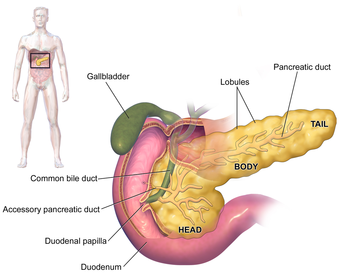
| Pancreas[1]
|
For patient information, click here
Editor-In-Chief: C. Michael Gibson, M.S., M.D. [1], Associate Editor(s)-in-Chief: ; Iqra Qamar M.D.[2]
Overview
Pancreatitis is an inflammatory disease of the pancreas characterized by reversible or irreversible changes in pancreatic structure and function leading to inflammation and fibrosis. The concept of pancreas and pancreatic duct was first described by Johannes Wirsung of Padua in 1642. Pancreatitis may be classified as acute pancreatitis, chronic pancreatitis, autoimmune pancreatitis, and hereditary pancreatitis. Common causes of pancreatitis may include gallstones, hypertriglyceridemia, alcohol, drugs, genetic, autoimmune, iatrogenic, trauma, infection, surgical causes, and obstruction. Acute pancreatitis usually presents with fever, sharp abdominal pain, nausea and vomiting. Patients with chronic pancreatitis present with dull abdominal pain, steatorrhea, pancreatic diabetes, nausea, weight loss, pseudocyst and pancreatic cancer.
Causes
Classification
Pancreatitis may be classified as:
Differential Diagnosis
Differentiating pancreatitis from other diseases on the basis of abdominal pain and weight loss:
Pancreatitis presents most commonly with abdominal pain. Pancreatitis must be differentiated from various disease which present with abdominal pain and weight loss such as peptic ulcer disease, pancreatic carcinoma, gastritis, and inflammatory bowel disease.
Abbreviations:
RUQ= Right upper quadrant of the abdomen, LUQ= Left upper quadrant, LLQ= Left lower quadrant, RLQ= Right lower quadrant, LFT= Liver function test, SIRS= Systemic inflammatory response syndrome, ERCP= Endoscopic retrograde cholangiopancreatography, IV= Intravenous, N= Normal, AMA= Anti mitochondrial antibodies, LDH= Lactate dehydrogenase, GI= Gastrointestinal, CXR= Chest X ray, IgA= Immunoglobulin A, IgG= Immunoglobulin G, IgM= Immunoglobulin M, CT= Computed tomography, PMN= Polymorphonuclear cells, ESR= Erythrocyte sedimentation rate, CRP= C-reactive protein, TS= Transferrin saturation, SF= Serum Ferritin, SMA= Superior mesenteric artery, SMV= Superior mesenteric vein, ECG= Electrocardiogram
| Disease
|
Clinical manifestations
|
Diagnosis
|
Comments
|
| Symptoms
|
Signs
|
| Abdominal Pain
|
Fever
|
Rigors and chills
|
Nausea or vomiting
|
Jaundice
|
Constipation
|
Diarrhea
|
Weight loss
|
GI bleeding
|
Hypo-
tension
|
Guarding
|
Rebound Tenderness
|
Bowel sounds
|
Lab Findings
|
Imaging
|
| Chronic pancreatitis
|
Epigastric
|
−
|
−
|
±
|
±
|
−
|
+
|
+
|
−
|
−
|
−
|
−
|
N
|
- Increased amylase / lipase
- Increased stool fat content
- Pancreatic function test
|
CT scan
- Calcification
- Pseudocyst
- Dilation of main pancreatic duct
|
- Predisposes to pancreatic cancer
|
| Pancreatic carcinoma
|
Epigastric
|
−
|
−
|
+
|
+
|
−
|
+
|
+
|
−
|
−
|
−
|
−
|
N
|
|
|
Skin manifestations may include:
|
| Peptic ulcer disease
|
Diffuse
|
±
|
−
|
+
|
−
|
−
|
−
|
+
|
|
Positive if perforated
|
Positive if perforated
|
Positive if perforated
|
N
|
- Ascitic fluid
- LDH > serum LDH
- Glucose < 50mg/dl
- Total protein > 1g/dl
|
|
|
| Disease
|
Abdominal Pain
|
Fever
|
Rigors and chills
|
Nausea or vomiting
|
Jaundice
|
Constipation
|
Diarrhea
|
Weight loss
|
GI bleeding
|
Hypo-
tension
|
Guarding
|
Rebound Tenderness
|
Bowel sounds
|
Lab Findings
|
Imaging
|
Comments
|
| Gastritis
|
Epigastric
|
±
|
−
|
+
|
−
|
−
|
−
|
Positive in chronic gastritis
|
+
|
−
|
−
|
−
|
N
|
|
|
|
| Gastric outlet obstruction
|
Epigastric
|
−
|
−
|
±
|
−
|
−
|
−
|
+
|
−
|
−
|
−
|
−
|
Hyperactive
|
|
|
|
| Gastroparesis
|
Epigastric
|
−
|
−
|
+
|
−
|
−
|
−
|
+
|
−
|
±
|
−
|
−
|
Hyperactive/hypoactive
|
- Hemoglobin
- Fasting plasma glucose
- Serum total protein, albumin, thyrotropin (TSH), and an antinuclear antibody (ANA) titer
- HbA1c
|
- Scintigraphic gastric emptying
|
- Succussion splash
- Single photon emission computed tomography (SPECT)
- Full thickness gastric and small intestinal biopsy
|
| Dumping syndrome
|
Lower and then diffuse
|
−
|
−
|
+
|
−
|
−
|
+
|
+
|
−
|
+
|
−
|
−
|
Hyperactive
|
- Glucose challenge test
- Hydrogen breath test
|
- Upper GI series
- Gastric emptying study
|
|
| Disease
|
Abdominal Pain
|
Fever
|
Rigors and chills
|
Nausea or vomiting
|
Jaundice
|
Constipation
|
Diarrhea
|
Weight loss
|
GI bleeding
|
Hypo-
tension
|
Guarding
|
Rebound Tenderness
|
Bowel sounds
|
Lab Findings
|
Imaging
|
Comments
|
| Inflammatory bowel disease
|
Diffuse
|
±
|
−
|
−
|
±
|
−
|
+
|
+
|
+
|
−
|
−
|
−
|
Normal or hyperactive
|
|
|
Extra intestinal findings:
|
| Irritable bowel syndrome
|
Diffuse
|
−
|
−
|
−
|
−
|
±
|
±
|
+
|
−
|
−
|
−
|
−
|
N
|
Normal
|
Normal
|
Symptomatic treatment
|
| Whipple's disease
|
Diffuse
|
±
|
−
|
−
|
±
|
−
|
+
|
+
|
−
|
±
|
−
|
−
|
N
|
|
Endoscopy is used to confirm diagnosis.
Images used to find complications
|
Extra intestinal findings:
|
| Disease
|
Abdominal Pain
|
Fever
|
Rigors and chills
|
Nausea or vomiting
|
Jaundice
|
Constipation
|
Diarrhea
|
Weight loss
|
GI bleeding
|
Hypo-
tension
|
Guarding
|
Rebound Tenderness
|
Bowel sounds
|
Lab Findings
|
Imaging
|
Comments
|
| Tropical sprue
|
Diffuse
|
+
|
−
|
−
|
−
|
−
|
+
|
+
|
−
|
−
|
−
|
−
|
N
|
|
Barium studies:
- Dilation and edema of mucosal folds
|
|
| Celiac disease
|
Diffuse
|
−
|
−
|
−
|
−
|
−
|
+
|
+
|
−
|
−
|
−
|
−
|
Hyperactive
|
|
US:
- Bull’s eye or target pattern
- Pseudokidney sign
|
|
| Colon carcinoma
|
Diffuse/localized
|
−
|
−
|
−
|
−
|
±
|
±
|
+
|
+
|
±
|
−
|
−
|
- Normal or hyperactive if obstruction present
|
- CBC
- Carcinoembryonic antigen (CEA)
|
- Colonoscopy
- Flexible sigmoidoscopy
- Barium enema
- CT colonography
|
- PILLCAM 2: A colon capsule for CRC screening may be used in patients with an incomplete colonoscopy who lacks obstruction
|
| Viral hepatitis
|
RUQ
|
+
|
−
|
+
|
+
|
−
|
Positive in Hep A and E
|
+
|
−
|
Positive in fulminant hepatitis
|
Positive in acute
|
+
|
N
|
- Abnormal LFTs
- Viral serology
|
|
- Hep A and E have fecal-oral route of transmission
- Hep B and C transmits via blood transfusion and sexual contact.
|
| Liver abscess
|
RUQ
|
+
|
+
|
+
|
+
|
−
|
±
|
+
|
−
|
+
|
+
|
±
|
Normal or hypoactive
|
|
|
|
| Hepatocellular carcinoma/Metastasis
|
RUQ
|
+
|
−
|
−
|
+
|
−
|
−
|
+
|
−
|
−
|
−
|
−
|
- Normal
- Hyperactive if obstruction present
|
|
|
Other symptoms:
|
| Disease
|
Abdominal Pain
|
Fever
|
Rigors and chills
|
Nausea or vomiting
|
Jaundice
|
Constipation
|
Diarrhea
|
Weight loss
|
GI bleeding
|
Hypo-
tension
|
Guarding
|
Rebound Tenderness
|
Bowel sounds
|
Lab Findings
|
Imaging
|
Comments
|
| Cirrhosis
|
RUQ
|
−
|
−
|
−
|
+
|
−
|
−
|
+
|
+
|
+
|
−
|
−
|
N
|
|
US
|
- Stigmata of liver disease
- Cruveilhier- Baumgarten murmur
|
| Small bowel obstruction
|
Diffuse
|
+
|
−
|
+
|
−
|
+
|
−
|
+
|
−
|
+
|
+
|
±
|
Hyperactive then absent
|
|
Abdominal X ray
- Dilated loops of bowel with air fluid levels
- Gasless abdomen
|
- "Target sign"– , indicative of intussusception
- Venous cut-off sign" – suggests thrombosis
|
| Mesenteric ischemia
|
Periumbilical
|
Positive if bowel becomes gangrenous
|
−
|
+
|
−
|
−
|
+
|
+
|
+
|
Positive if bowel becomes gangrenous
|
Positive if bowel becomes gangrenous
|
−
|
Hyperactive to absent
|
|
CT angiography
|
- Also known as abdominal angina that worsens with eating
|
| Acute ischemic colitis
|
Diffuse
|
+
|
±
|
+
|
−
|
−
|
+
|
+
|
+
|
+
|
+
|
+
|
Hyperactive then absent
|
|
Abdominal x-ray
- Distension and pneumatosis
CT scan
- Double halo appearance, thumbprinting
- Thickening of bowel
|
|
| Ruptured abdominal aortic aneurysm
|
Diffuse
|
±
|
−
|
+
|
−
|
−
|
−
|
+
|
+
|
+
|
−
|
−
|
N
|
|
- Focused Assessment with Sonography in Trauma (FAST)
|
|
| Pleural empyema
|
RUQ/Epigastric
|
+
|
±
|
−
|
−
|
−
|
−
|
+
|
−
|
−
|
−
|
−
|
N
|
|
Chest X-ray
|
Physical examination
|
|
Template:WikiDoc Sources
