Leukocytosis
|
WikiDoc Resources for Leukocytosis |
|
Articles |
|---|
|
Most recent articles on Leukocytosis Most cited articles on Leukocytosis |
|
Media |
|
Powerpoint slides on Leukocytosis |
|
Evidence Based Medicine |
|
Clinical Trials |
|
Ongoing Trials on Leukocytosis at Clinical Trials.gov Clinical Trials on Leukocytosis at Google
|
|
Guidelines / Policies / Govt |
|
US National Guidelines Clearinghouse on Leukocytosis
|
|
Books |
|
News |
|
Commentary |
|
Definitions |
|
Patient Resources / Community |
|
Patient resources on Leukocytosis Discussion groups on Leukocytosis Patient Handouts on Leukocytosis Directions to Hospitals Treating Leukocytosis Risk calculators and risk factors for Leukocytosis
|
|
Healthcare Provider Resources |
|
Causes & Risk Factors for Leukocytosis |
|
Continuing Medical Education (CME) |
|
International |
|
|
|
Business |
|
Experimental / Informatics |
Editor-In-Chief: C. Michael Gibson, M.S., M.D. [1] Associate Editor(s)-in-Chief: Maria Fernanda Villarreal, M.D. [2] Lakshmi Gopalakrishnan, M.B.B.S. [3]
Synonyms and keywords: Elevated white blood cell count; Right-shift leukocytosis; Left-leukocytosis
Overview
Leukocytosis is an elevation of the white blood cell count above the normal range (greater than 11,000 per mm3). Leukocytosis is frequently a sign of an inflammatory response, most commonly the result of infection, but may also occur following certain parasitic infections, bone tumors, strenuous exercise, emotional stress, pregnancy, anesthesia, and epinephrine administration. Leukocytosis may be classified into 5 subtypes: neutrophilia (most common), lymphocytosis, monocytosis, eosinophilia, and basophilia. Other classification, include: Left shift or right shift leucocytosis. The pathogenesis of leukocytosis is characterized by the increase of leukocytes (primarily neutrophils), followed by the proliferation and release of granulocyte and monocyte precursors in the bone marrow which is stimulated by several products of inflammation including C3a and G-CSF.[1]
Historical Perspective
- Leukocytosis was first discovered by Paul Kautchakoff, in 1846.[1]
Classification
- Leukocytosis may be classified into 5 subtypes:[2]
- Neutrophilia (most common)
- Lymphocytosis
- Monocytosis
- Eosinophilia
- Basophilia.
- Leukocytosis may also be classified into 2 groups:[2]
- Left shift (most common)
- Immature leukocytes increase
- Proliferation and release of granulocyte and monocyte precursors in the bone marrow
- Usually stimulated by several products of inflammation including C3a and G-CSF
- Right shift
- Reduced count or lack of "young neutrophils"
- Associated with the presence of "giant neutrophils
- Other variant of leukocytosis is the leukemoid reaction.
- The image below demonstrates a graphic figure that illustrates hematopoietic growth factors in leukocytosis.[3]
-
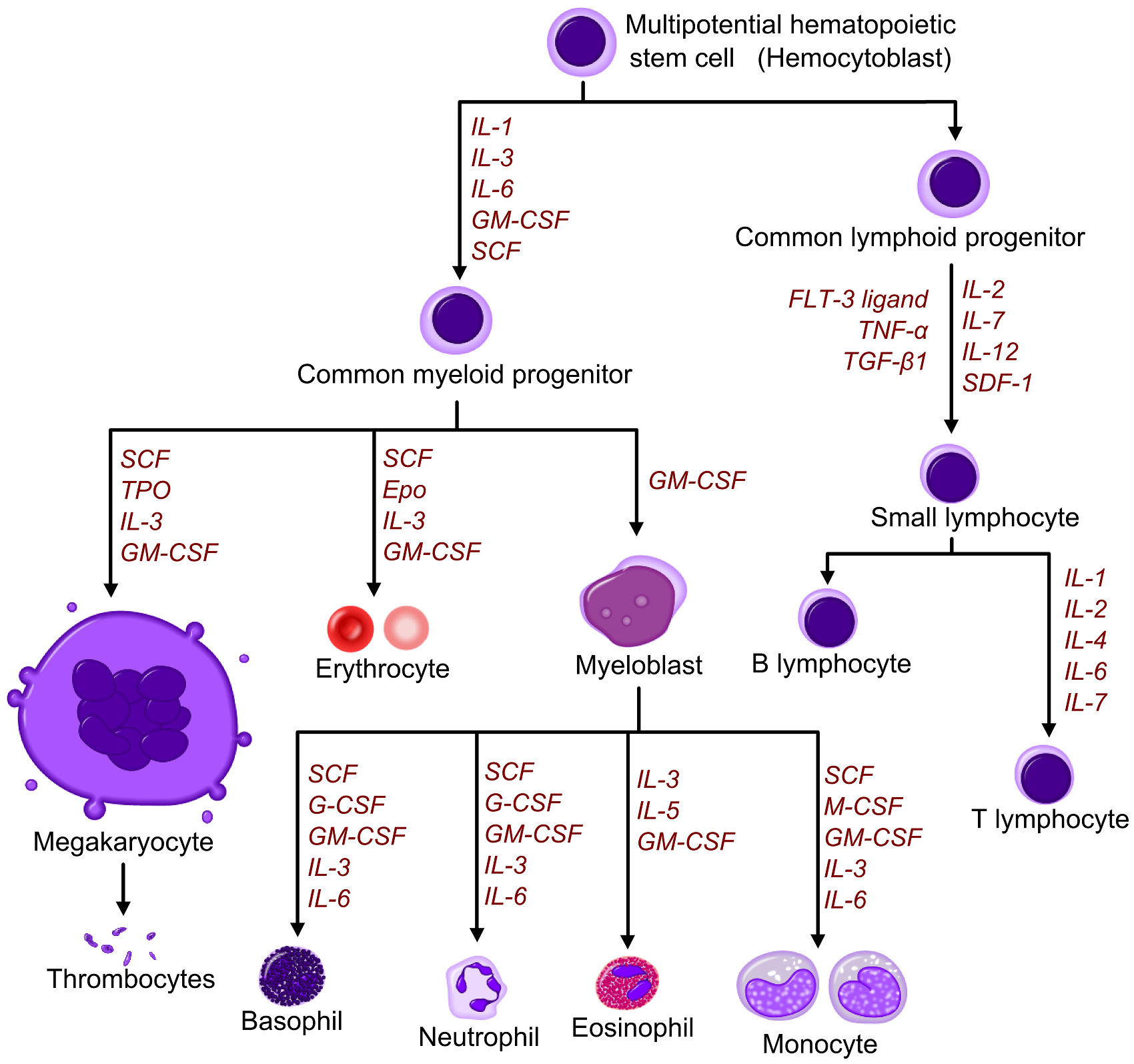
Hematopoietic growth factors in leukocytosis
Courtesy of Wikipedia

Pathophysiology
- The pathogenesis of leukocytosis is characterized by:[2]
- An increased release of leukocytes from bone marrow storage pools
- Decreased margination of leukocytes onto vessel walls
- Decreased extravasation of leukocytes from the vessels into tissues
- Increase in number of precursor cells in the marrow
Causes
- To see a comprehensive list of all causes of leukocytosis, please click here
| Causes of leukocytosis | ||||
|---|---|---|---|---|
| Neutrophilic leukocytosis (neutrophilia) |
||||
| Eosinophilic leukocytosis (eosinophilia) |
| |||
| Basophilic leukocytosis Basophilia |
||||
| Monocytosis | ||||
| Lymphocytosis |
| |||
Epidemiology and Demographics
- Leukocytosis is very common.[2]
Age
- Patients of all age groups may develop leukocytosis.
- Normal white blood count differential changes with age.
- Leukocytosis in neonates is more common, compared to children and adults.[2]
Gender
- Leukocytosis affects men and women equally.
Race
- There is no racial predilection for leukocytosis.
Risk Factors
- Common risk factors in the development of leukocytosis, include:[2]
- Physiologic processes (eg, stress, exercise, pregnancy)
- Drugs (e.g.corticosteroids, lithium, beta agonists)
- Trauma
- Stress
Natural History, Complications and Prognosis
- The majority of patients with leukocytosis are initially symptomatic.[3]
- Early clinical features, include:[3]
- Fever
- Hyperhidrosis
- Fatigue
- Common complications of leukocytosis, include:[3]
- Tumor lysis syndrome
- Disseminated intravascular coagulopathy
- Acute respiratory failure
- Pulmonary hemorrhage
- CNS infarction
- Splenic infarction
- Myocardial ischemia
- Renal failure
- Prognosis is generally depends on the underlying etiologies.[3]
Diagnosis
Symptoms
- Leukocytosis is usually symptomatic.
- Symptoms of leukocytosis are often unspecific, such as:[3]
- Weight loss
- Fevers of unknown origin
- Hyperhidrosis
- Chronic pain
- Fatigue
- Dyspnea
- Malaise
- Obtain history of the following:
- Clinical features
- Duration (e.g. days, weeks, months)
- Remainder of complete blood count
Laboratory Findings
- Laboratory findings consistent with the diagnosis of leukocytosis, include:[3]
- White blood cell count above the normal range
- Greater than 11,000 per mm3
Differentiating Leukocytosis from Other Diseases
| Category | Condition | Etiology | Mechanism | Congenital | Acquried | Clinical manifestations | Para−clinical findings | Gold standard | Associated findings | ||||||||||||||||||||
|---|---|---|---|---|---|---|---|---|---|---|---|---|---|---|---|---|---|---|---|---|---|---|---|---|---|---|---|---|---|
| Demography | History | Symptoms | Signs | ||||||||||||||||||||||||||
| Lab Findings | |||||||||||||||||||||||||||||
| Physiologic | Increased bone marrow production | Demargination of peripheral blood neutrophils | Appearance | Fever | Abdominal pain | BP | Asplenia | Hepatosplenomegaly | Lymphadenopathy | Joint involvement | Other | CBC | PBS | Bone marrow exam | ESR/CRP | BUN/Cr | LFT | ||||||||||||
| Autonomous | Reactive | WBC | HB | Plt | |||||||||||||||||||||||||
| Hematologic | Hereditary neutrophilia[4] | − | + | − | − | + | − | Rare autosomal dominant genetic disorder |
|
Normal | − | − | Nl | − | + | − | − | − | ↑ | Nl | Nl |
|
Nl | Nl | Nl | Nl | Molecular testing |
| |
| Myeloproliferative neoplasms[5] |
|
− | + | − | − | + | + | Elderly | Exposure to | ± | + | Nl | − | + | − | − | ↑/↓ | ↓ | ↑/↓ |
|
|
↑ | Nl | Nl | Bone marrow examination + clinical manifestation |
| |||
| Polycythemia vera[6] |
|
− | + | − | − | + | − | Mean age >60 years old |
|
− | − | ↑ | − | + | + | − |
|
Nl to ↑ | ↑ | ↑ |
|
|
Nl | Nl | Nl | Bone marrow examination + clinical manifestation | |||
| Microangiopathic hemolytic anemia (MAHA)[7] | − | + | − | − | + | + | Any |
|
+ | + | ↓ | − | + | − | − | ↑ | ↓ | ↑ |
|
NA | ↑ | ↑ | ↑ | Bone marrow examination + clinical manifestation |
| ||||
| Leukoerythroblastosis[8] |
|
− | + | − | − | − | + | Any | − | + | Nl | − | + | − | − |
|
↑ | ↓ | ↓ |
|
|
↑ | Nl | ↑ | Bone marrow biopsy |
| |||
| Immunology/
Rheumatology |
Condition | Etiology | Physiologic | Autonomous increased bone marrow production | Reactive increased bone marrow production | Demargination of peripheral blood neutrophils | Congenital | Acquried | Demography | History | Appearance | Fever | Abdominal pain | BP | Asplenia | Hepatosplenomegaly | Lymphadenopathy | Joint involvement | Other signs | WBC | HB | Plt | PBS | Bone marrow exam | ESR/CRP | BUN/Cr | LFT | Gold standard | Associated findings |
| Leukocyte adhesion deficiency[9] |
|
− | + | − | − | + | − | Rare autosomal recessive, LAD II more in Middle East and Brazil |
|
|
+ | − | Nl | − | − | − | − |
|
↑ | ↓ | ↓/↑ |
|
|
Nl | Nl | Nl | Flow cytometry |
| |
| Cryopyrin-associated periodic syndromes[10] |
|
− | + | − | − | + | − | Autosomal dominant autoinflammatory syndrome |
|
|
+ | − | Nl | − | − | − | + | ↑ | ↓ | ↓/↑ |
|
|
↑ | Nl | ↑ | Genetic tests | |||
| Rheumatoid arthritis[11][12] |
|
− | + | − | − | − | + | Any, more in young women, between 30-60 years old |
|
|
+ | − | Nl | − | − | − | + |
|
↑ | ↓ | ↑ |
|
NA | ↑ | Nl | Nl | Clinical manifestation + positive anti-CCP antibodies |
| |
| Juvenile onset rheumatoid arthritis[13] |
|
− | + | − | − | − | + | Children under the age of 16 |
|
|
+ | − | Nl | − | − | + | + |
|
↑ | ↓ | ↑ |
|
NA | ↑ | Nl | Nl | Clinical manifestation + laboratory findings |
| |
| Adult Still's disease |
|
− | + | − | − | − | + | Rare autoimmune disease | NA | + | − | Nl | − | − | + | + | ↑ | ↓ | ↑ |
|
NA | ↑ | Nl | Nl | Diagnosis of exclusion |
| |||
| Kawasaki disease |
|
− | + | − | − | − | + | Autoimmune disease, more in Asian ethnicity boys | NA |
|
+ | + | Nl | − | − | + | + |
|
↑ | ↓ | ↑ |
|
NA | ↑ | Nl | Nl | Diagnostic criteria |
| |
| IBD |
|
− | + | − | − | − | + | Autoimmune disease, more in young |
|
|
+ | + | Nl | − | + | + | + |
|
↑ | ↓ | ↑ |
|
NA | ↑ | Nl | Nl | Colonoscopy and biopsy |
| |
| Sarcoidosis |
|
− | + | − | − | − | + | Autoimmune disease, more in young African American women |
|
|
+ | + | Nl | − | + | +
Bilateral hilar |
+ |
|
↑ | ↓ | ↑ |
|
NA | ↑ | Nl | Nl | Diagnosis of exclusion |
| |
| Chronic hepatitis |
|
− | + | − | − | − | + | Elderly |
|
|
+ | + | ↓ | − | + | + | + |
|
↑ | ↓ | ↑ |
|
NA | ↑ | ↑ | ↑ | Liver biopsy |
| |
| Sweet syndrome |
|
− | + | − | − | − | + | Rare |
|
|
+ | + | Nl | − | − | + | + |
|
↑ | ↓ | ↑ |
|
|
↑ | Nl | Nl | Diagnostic criteria |
| |
| Acute gout |
|
− | + | − | − | − | + | Older males |
|
|
+ | − | − | − | − | + |
|
↑ | ↓ | ↑ |
|
NA | ↑ | ↑ | Nl | Clinical manifestation |
| ||
| Medication | Condition | Etiology | Physiologic | Autonomous increased bone marrow production | Reactive increased bone marrow production | Demargination of peripheral blood neutrophils | Congenital | Acquried | Demography | History | Appearance | Fever | Abdominal pain | BP | Asplenia | Hepatosplenomegaly | Lymphadenopathy | Joint involvement | Other signs | WBC | HB | Plt | PBS | Bone marrow exam | ESR/CRP | BUN/Cr | LFT | Gold standard | Associated findings |
| Steroid |
|
− | − | + | + | − | + | Elderly |
|
|
− | + | Nl to ↓ | − | − | − | − | ↑ | Nl to ↓ | ↑ |
|
|
↑ | ↑ | Nl | Clinical manifestation + history of drug consumption |
| ||
| Myeloid growth factors | |||||||||||||||||||||||||||||
| Lithium | |||||||||||||||||||||||||||||
| Cytokines | |||||||||||||||||||||||||||||
| Catecholamines |
|
− | − | + | + | − | + | Any |
|
|
− | − | Nl to ↓ | − | − | − | − | ↑ | ↑ | ↑ |
|
|
↑ | Nl | Nl | Clinical manifestation + history of drug consumption |
| ||
| ATRA | |||||||||||||||||||||||||||||
| Other | Condition | Etiology | Physiologic | Autonomous increased bone marrow production | Reactive increased bone marrow production | Demargination of peripheral blood neutrophils | Congenital | Acquried | Demography | History | Appearance | Fever | Abdominal pain | BP | Asplenia | Hepatosplenomegaly | Lymphadenopathy | Joint involvement | Other signs | WBC | HB | Plt | PBS | Bone marrow exam | ESR/CRP | BUN/Cr | LFT | Gold standard | Associated findings |
| Infections | Infection | - | + | + | + | - | + | Any | |||||||||||||||||||||
| Allergy |
|
Any |
|
|
− | − | Nl to ↓ | − | − | − | − | ↑ | ↑ | ↑ |
|
|
↑ | Nl | Nl | Clinical manifestation | − | ||||||||
| Post splenectomy |
|
− | − | + | + | − | + | Any |
|
|
± | − | Nl | + | − | − | − | − | ↑ | ↓ | ↑ |
|
|
↑ | Nl | Nl | Clinical manifestation |
| |
| Down syndrome | |||||||||||||||||||||||||||||
| Cigarette smoking | |||||||||||||||||||||||||||||
| Stress/exercise | Athlete |
|
|
− | − | Nl | − | − | − | − | − | ↑ | ↑ | ↑ |
|
|
↑ | Nl | Nl | Clinical manifestation | − | ||||||||
| Infancy | |||||||||||||||||||||||||||||
| Pregnancy | |||||||||||||||||||||||||||||
| Platelet clumping | Spurious | ||||||||||||||||||||||||||||
| Mixed cryoglobulinemia | Spurious | ||||||||||||||||||||||||||||
| Category | Condition | Etiology | Physiologic | Autonomous increased bone marrow production | Reactive increased bone marrow production | Demargination of peripheral blood neutrophils | Congenital | Acquried | Demography | History | Appearance | Fever | Abdominal pain | BP | Asplenia | Hepatosplenomegaly | Lymphadenopathy | Joint involvement | Other signs | WBC | HB | Plt | PBS | Bone marrow exam | ESR/CRP | BUN/Cr | LFT | Gold standard | Associated findings |
Treatment
Medical Therapy
- The treatment for leukocytosis will depend on the underlying condition.[3]
References
- ↑ 1.0 1.1 Chabot-Richards DS, George TI (2014). "Leukocytosis". Int J Lab Hematol. 36 (3): 279–88. doi:10.1111/ijlh.12212. PMID 24750674.
- ↑ 2.0 2.1 2.2 2.3 2.4 2.5 Abramson N, Melton B (2000). "Leukocytosis: basics of clinical assessment". Am Fam Physician. 62 (9): 2053–60. PMID 11087187.
- ↑ 3.0 3.1 3.2 3.3 3.4 3.5 3.6 3.7 Leukocytosis. Wikipedia. https://en.wikipedia.org/wiki/Leukocytosis Accessed on May 23, 2016
- ↑ Herring, William Benjamin; Smith, Laurin Gresham; Walker, Richard Isley; Herion, John Carroll (1974). "Hereditary neutrophilia". The American Journal of Medicine. 56 (5): 729–734. doi:10.1016/0002-9343(74)90642-1. ISSN 0002-9343.
- ↑ Tefferi A (February 2010). "Leukocytosis as a risk factor for thrombosis in myeloproliferative neoplasms-biologically plausible but clinically uncertain". Am. J. Hematol. 85 (2): 93–4. doi:10.1002/ajh.21614. PMID 20052751.
- ↑ Boiocchi L, Gianelli U, Iurlo A, Fend F, Bonzheim I, Cattaneo D, Knowles DM, Orazi A (November 2015). "Neutrophilic leukocytosis in advanced stage polycythemia vera: hematopathologic features and prognostic implications". Mod. Pathol. 28 (11): 1448–57. doi:10.1038/modpathol.2015.100. PMID 26336886.
- ↑ Morton JM, George JN (June 2016). "Microangiopathic Hemolytic Anemia and Thrombocytopenia in Patients With Cancer". J Oncol Pract. 12 (6): 523–30. doi:10.1200/JOP.2016.012096. PMID 27288467.
- ↑ Canbolat Ayhan A, Timur C, Ayhan Y, Kes G (June 2014). "Leukoerythroblastosis Mimicking Leukemia: A case report". Iran J Pediatr. 24 (3): 332–3. PMC 4276592. PMID 25562031.
- ↑ Levy-Mendelovich S, Rechavi E, Abuzaitoun O, Vernitsky H, Simon AJ, Lev A, Somech R (April 2016). "Highlighting the problematic reliance on CD18 for diagnosing leukocyte adhesion deficiency type 1". Immunol. Res. 64 (2): 476–82. doi:10.1007/s12026-015-8706-5. PMID 26434744.
- ↑ Labrousse M, Kevorkian-Verguet C, Boursier G, Rowczenio D, Maurier F, Lazaro E, Aggarwal M, Lemelle I, Mura T, Belot A, Touitou I, Sarrabay G (September 2018). "Mosaicism in autoinflammatory diseases: Cryopyrin-associated periodic syndromes (CAPS) and beyond. A systematic review". Crit Rev Clin Lab Sci. 55 (6): 432–442. doi:10.1080/10408363.2018.1488805. PMID 30035647.
- ↑ Scott DL, Wolfe F, Huizinga TW (September 2010). "Rheumatoid arthritis". Lancet. 376 (9746): 1094–108. doi:10.1016/S0140-6736(10)60826-4. PMID 20870100.
- ↑ Glant TT, Mikecz K, Rauch TA (February 2014). "Epigenetics in the pathogenesis of rheumatoid arthritis". BMC Med. 12: 35. doi:10.1186/1741-7015-12-35. PMC 3936819. PMID 24568138.
- ↑ Naz S, Mushtaq A, Rehman S, Bari A, Maqsud A, Khan MZ, Ahmad TM (June 2013). "Juvenile rheumatoid arthritis". J Coll Physicians Surg Pak. 23 (6): 409–12. doi:06.2013/JCPSP.409412 Check
|doi=value (help). PMID 23763801.