Leiomyoma: Difference between revisions
Shanshan Cen (talk | contribs) No edit summary |
Shanshan Cen (talk | contribs) No edit summary |
||
| Line 1: | Line 1: | ||
__NOTOC__ | __NOTOC__ | ||
{{Leiomyoma}} | {{Leiomyoma}} | ||
{{CMG}}; {{AE}} {{CZ}} {{SC}} | {{CMG}}; {{AE}} {{CZ}} {{SC}} | ||
{{SK}} | {{SK}} | ||
==Overview== | |||
== | |||
==Pathophysiology== | ==Pathophysiology== | ||
| Line 126: | Line 97: | ||
</gallery> | </gallery> | ||
==ultrasound== | ==ultrasound== | ||
Revision as of 15:47, 9 December 2015
Editor-In-Chief: C. Michael Gibson, M.S., M.D. [1]; Associate Editor(s)-in-Chief: Cafer Zorkun, M.D., Ph.D. [2] Shanshan Cen, M.D. [3]
Synonyms and keywords:
Overview
Pathophysiology
Uterine Leiomyomata
Uterine fibroids are leiomyomata of the uterine smooth muscle. As other leiomyomata, they are benign, but may lead to excessive menstrual bleeding (menorrhagia), often cause anemia and may lead to infertility. Enucleation is removal of fibroids without removing the uterus (hysterectomy), which is also commonly performed. Laser surgery (called myolysis) is increasingly used, and provides a viable alternative to surgery.
Uterine leiomyomas originate in the myometrium and are classified by location:
- Submucosal – lie just beneath the endometrium.
- Intramural – lie within the uterine wall.
- Subserous – lies at the serosal surface of the uterus or may bulge out from the myometrium and can become pedunculated.
Estrogen and progesterone usually stimulate their growth, and hormone suppression may hence decrease their size.
Esophageal
They are also the most common benign esophageal tumour, though this accounts for less than 1% of esophageal neoplasms. The remainder consists mainly of carcinomas. Although the vast majority of benign esophageal tumors are clinically silent and go undetected, large or strategically located tumors may become symptomatic. [1]
Leiomyoma of Jejunum
Leiomyoma is the most common benign tumor of small bowel. Approximately 50% of cases are found in the jejunum, followed by the ileum in 31% of cases. Almost one half of all lesions are less than 5 centimeters. [2]
Gross Pathology
-
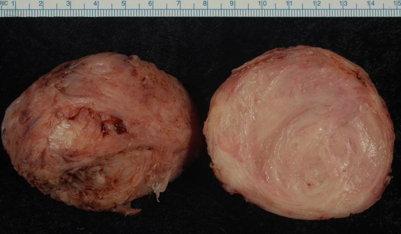
Leiomyoma enucleated from a uterus. External surface on left; cut surface on right
-

A large, solitary leiomyoma in the uterus, distoring the endometrial cavity into a Y shape by splaying and pressing it downwards.
(Image courtesy of Ed Uthman, MD)


epidemiology and demographics
The disease is 5 times more common in Afro-American females.
CT
CT images demonstrate a right broad ligament leiomyoma


MRI
- Differential diagnosis at MR imaging includes adenomyosis, solid adnexal mass, focal myometrial contraction, and uterine leiomyosarcoma.
- Nondegenerated uterine leiomyomas have a typical appearance at MR imaging: well-circumscribed masses of homogeneously decreased signal intensity compared with that of the outer myometrium on T2-weighted images.
- Degenerated leiomyomas have variable appearances on T2-weighted images and contrast-enhanced images.
Patient #1: MR images demonstrate large degenerating leiomyomas


Patient #2: MR images demonstrate a leiomyoma prolapsing into the endometrial canal


ultrasound
Diagnosis depends on position of the myomas. Traditional transabdominal or transvaginal ultrasonogram can detect large submucosal and transmural myomas. For submucosal myomas saline infusion sonography is the best.
Other Imaging Findings
Hysterosalpingogram(HSG)
Hysterosalpingogram(HSG) reveals a submucosal leiomyoma


medical therapy
GnRH analogues for 3-6 months are used to reduce the size of the myomas. It usually reduce the size by 60% to 70%. But once the medication is stopped the myomas will grow back.
surgery
Myomectomy is a choice to remove myomas. It is usually done when the client wants to preserve their fertility. Total Abdominal or Vaginal hysterectomy with Bilateral Salpingo-oophorectomy is the definitive treatment.
- ↑ James C. Chou, MD & Frank G. Gress, MD. "Benign Esophageal Tumors". Esophageal Cancer Overview (Cancer of the Esophagus). Retrieved 2007-03-21. Unknown parameter
|publsiher=ignored (|publisher=suggested) (help) - ↑ By Michael P. Buetow, M.D. "Leiomyoma of Jejunum". Retrieved 2007-03-21. Unknown parameter
|publsiher=ignored (|publisher=suggested) (help)