Uterus
|
WikiDoc Resources for Uterus |
|
Articles |
|---|
|
Media |
|
Evidence Based Medicine |
|
Clinical Trials |
|
Ongoing Trials on Uterus at Clinical Trials.gov Clinical Trials on Uterus at Google
|
|
Guidelines / Policies / Govt |
|
US National Guidelines Clearinghouse on Uterus
|
|
Books |
|
News |
|
Commentary |
|
Definitions |
|
Patient Resources / Community |
|
Directions to Hospitals Treating Uterus Risk calculators and risk factors for Uterus
|
|
Healthcare Provider Resources |
|
Continuing Medical Education (CME) |
|
International |
|
|
|
Business |
|
Experimental / Informatics |
Editor-In-Chief: C. Michael Gibson, M.S., M.D. [1]
Overview
The uterus or womb is the major female reproductive organ of most mammals, including humans. One end, the cervix, opens into the vagina; the other is connected on both sides to the fallopian tubes. The term uterus is commonly used within the medical and related professions, whilst womb is in more common usage. The plural of uterus is uteri.
Function
The main function of the uterus is to accept a fertilized ovum which becomes implanted into the endometrium, and derives nourishment from blood vessels which develop exclusively for this purpose. The fertilized ovum becomes an embryo, develops into a fetus and gestates until childbirth. Due to anatomical barriers such as the pelvis, the uterus is pushed partially into the abdomen due to its expansion during pregnancy. Even in pregnancy the mass of a human uterus amounts to only about a kilogram (2.2 pounds).
Forms in mammals
In mammals, the four main forms in which it is found are:
- Bipartite
- Found in ruminants (cattle, goats, sheep, camels, llamas, giraffes, bison, buffalo, deer, etc.).
- Bicornuate
- Found in pigs, cats, and dogs.
- Simplex
- Found in humans, other primates and horses.
- Duplex
- Found in rodents (such as mice, rats and guinea pigs), marsupials and lagomorpha (rabbits and hares).
Anatomy
The uterus is located inside the pelvis immediately dorsal (and usually somewhat rostral) to the urinary bladder and ventral to the rectum. Outside of pregnancy, its size in humans is several centimeters in diameter.
Regions
From outside to inside, the path to the uterus is as follows:
Layers
The layers, from innermost to outermost, are as follows:
- Endometrium
- The lining of the uterine cavity is called the "endometrium." In most mammals, including humans, the endometrium builds a lining periodically which, if no pregnancy occurs, is shed or reabsorbed. Shedding of the endometrial lining in humans is responsible for menstrual bleeding (known colloquially as a woman's "period") throughout the fertile years of a female and for some time beyond. In other mammals there may be cycles set as widely apart as six months or as frequently as a few days.
- Myometrium
- The uterus mostly consists of smooth muscle, known as "myometrium." The innermost layer of myometrium is known as the junctional zone, which becomes thickened in adenomyosis.
- Perimetrium
- The loose surrounding tissue is called the "perimetrium."
- Peritoneum
- The uterus is surrounded by "peritoneum."
Support
The uterus is primarily supported by the pelvic diaphragm and the urogenital diaphragm. Secondarily, it is supported by ligaments and the peritoneum (broad ligament of the uterus) [1]
Major ligaments
It is held in place by several peritoneal ligaments, of which the following are the most important (there are two of each):
| Name | From | To |
|---|---|---|
| uterosacral ligament | the posterior cervix | the sacrum of pelvis |
| cardinal ligaments | the side of the cervix | the ischial spines |
| pubocervical ligament [1] |
Other named ligaments near the uterus, i.e. the broad ligament, the round ligament, the suspensory ligament of the ovary, the infundibulopelvic ligament, have no role in the support of the uterus.
Position
Under normal circumstances the uterus is both "anteflexed" and "anteverted." The meaning of these terms are described below:
| Distinction | More common | Less common |
|---|---|---|
| Position tipped | "anteverted": tipped forward | "retroverted": tipped backwards |
| Position of fundus | "anteflexed": the fundus is pointing forward relative to the cervix | "retroflexed": the fundus is pointing backwards |
Development
The bilateral Müllerian ducts form during early fetal life. In males, MIF secreted from the testes leads to their regression. In females these ducts give rise to the Fallopian tubes and the uterus. In humans the lower segments of the two ducts fuse to form a single uterus, however, in cases of uterine malformations this development may be disturbed. The different uterine forms in various mammals are due to various degrees of fusion of the two Müllerian ducts.
Pathology
Some pathological states include:
- Prolapse of the uterus
- Carcinoma of the cervix – malignant neoplasm
- Carcinoma of the uterus – malignant neoplasm
- Ectopic pregnancy
- Fibroids – benign neoplasms
- Adenomyosis – ectopic growth of endometrial tissue within the myometrium
- Pyometra
- Uterine malformation
- Uterine Didelphys – split or doubled vagina/uterus
- Retroverted uterus
- Rokitansky syndrome
- Myoma
Additional images
CT images of a pregnant patient thought to have appendicitis. Benefits of CT scan felt to outweigh the risks. See the gravid uterus


Additional images
-
Schematic frontal view of female anatomy
-
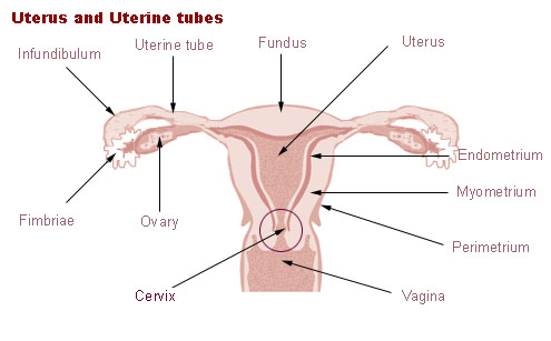 Uterus and uterine tubes.
 -

-
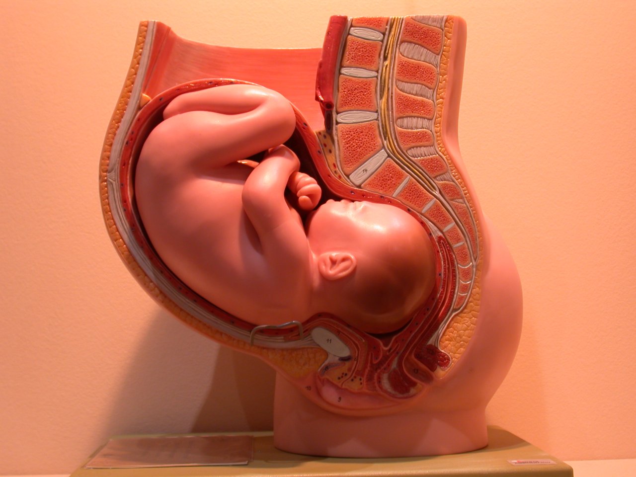 Anatomical model of a human pregnancy
 -
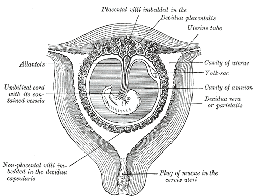 Sectional plan of the gravid uterus in the third and fourth month.
 -
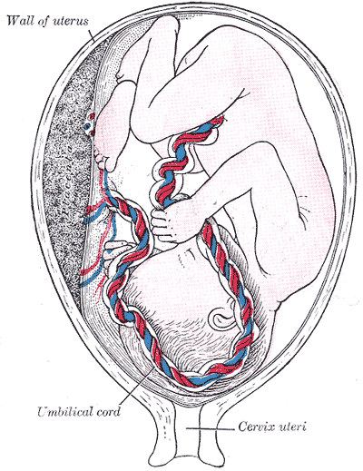 Fetus in utero, between fifth and sixth months.
 -
 Vessels of the uterus and its appendages, rear view.
 -
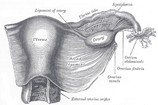 Uterus and right broad ligament, seen from behind.
 -
 Female pelvis and its contents, seen from above and in front.
 -
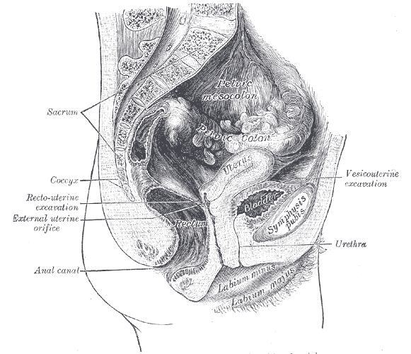
 Sagittal section of the lower part of a female trunk, right segment.
 -
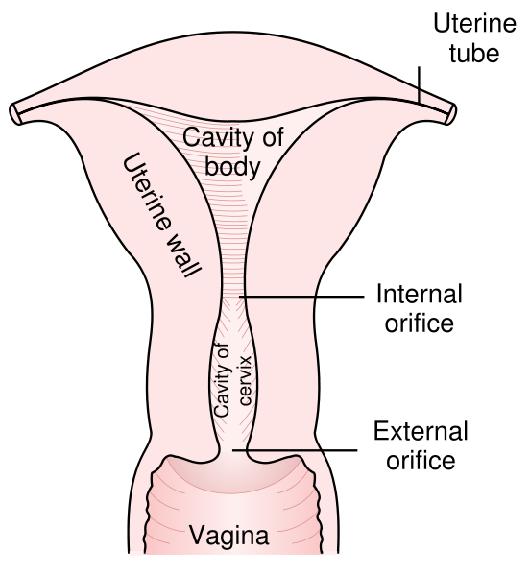 Posterior half of uterus and upper part of vagina.
 -
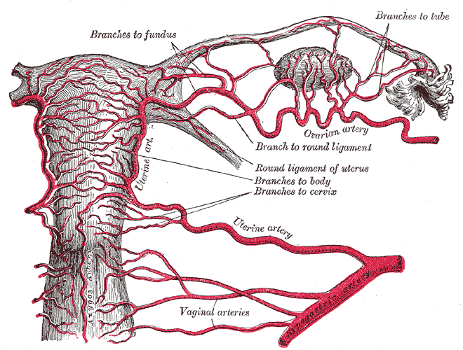 The arteries of the internal organs of generation of the female, seen from behind.
 -
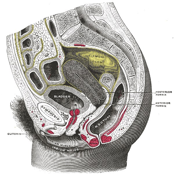
 Median sagittal section of female pelvis.


References
- ↑ 1.0 1.1 The Pelvis University College Cork
See also
- WikiSaurus:womb – the WikiSaurus list of synonyms and slang words for the womb in many languages
- Myometrium
- Ovum
- Ovary
- Menopause
- Vulva
- Vagina
- Penis
- Uterine glands
External links
- Template:GraySubject
- Illustration at wku.edu
- Template:SUNYAnatomyLabs - "The Female Pelvis: Organs in the Female Pelvis in situ"
Template:Female reproductive system
ar:رحم
zh-min-nan:Seⁿ-kiáⁿ-tē
bs:Materica
bg:Матка
cs:Děloha
cy:Croth
da:Livmoder
pdc:Mudder (Uterus)
de:Gebärmutter
dv:ރަހިމު
et:Emakas
eo:Utero
gd:Machlag
gl:Útero
hi:गर्भ
id:Rahim (anatomi)
it:Utero
he:רחם
ku:Malzarok
la:Uterus
lt:Gimda
hu:Méh (szerv)
ml:ഗര്ഭപാത്രം
mr:गर्भाशय
nl:Baarmoeder
no:Livmor
oc:Utèr
qu:Kisma
simple:Uterus
sk:Maternica
sl:Maternica
sr:Материца
su:Rahim (anatomi)
fi:Kohtu
sv:Livmoder
ta:கருப்பை
te:గర్భాశయం
uk:Матка
yi:רחם