Peritoneum
Editor-In-Chief: C. Michael Gibson, M.S., M.D. [1]
Overview
|
WikiDoc Resources for Peritoneum |
|
Articles |
|---|
|
Most recent articles on Peritoneum |
|
Media |
|
Evidence Based Medicine |
|
Clinical Trials |
|
Ongoing Trials on Peritoneum at Clinical Trials.gov Clinical Trials on Peritoneum at Google
|
|
Guidelines / Policies / Govt |
|
US National Guidelines Clearinghouse on Peritoneum
|
|
Books |
|
News |
|
Commentary |
|
Definitions |
|
Patient Resources / Community |
|
Patient resources on Peritoneum Discussion groups on Peritoneum Patient Handouts on Peritoneum Directions to Hospitals Treating Peritoneum Risk calculators and risk factors for Peritoneum
|
|
Healthcare Provider Resources |
|
Causes & Risk Factors for Peritoneum |
|
Continuing Medical Education (CME) |
|
International |
|
|
|
Business |
|
Experimental / Informatics |
In higher vertebrates, the peritoneum is the serous membrane that forms the lining of the abdominal cavity — it covers most of the intra-abdominal organs. It is composed of a layer of mesothelium supported by a thin layer of connective tissue.
The peritoneum both supports the abdominal organs and serves as a conduit for their blood and lymph vessels and nerves.
Structure
Layers
The abdominal cavity (the space bounded by the vertebrae, abdominal muscles, diaphragm and pelvic floor) should not be confused with the intraperitoneal space (located within the abdominal cavity, but wrapped in peritoneum). For example, a kidney is inside the abdominal cavity, but is retroperitoneal.
Although they ultimately form one continuous sheet, two types or layers of peritoneum and a potential space between them are referenced:
- The outer layer, called the parietal peritoneum, is attached to the abdominal wall.
- The inner layer, the visceral peritoneum, is wrapped around the internal organs that are located inside the intraperitoneal cavity.
- The potential space between these two layers is the peritoneal cavity; it is filled with a small amount (about 50 ml) of slippery serous fluid that allows the two layers to slide freely over each other.
- The term mesentery is often used to refer to a double layer of visceral peritoneum. There are often blood vessels, nerves, and other structures between these layers. It should be noted that the space between these two layers is technically outside of the peritoneal sac, and thus not in the peritoneal cavity.
Subdivisions
There are two main regions of the peritoneum, connected by the epiploic foramen:
- the greater sac (or general cavity of the abdomen), represented in red in the diagrams above.
- the lesser sac (or omental bursa), represented in blue. The lesser sac is divided into two "omenta":
- The lesser omentum (or gastrohepatic) is attached to the lesser curvature of the stomach and the liver.
- The greater omentum (or gastrocolic) hangs from the greater curve of the stomach and loops down in front of the intestines before curving back upwards to attach to the transverse colon. In effect it is draped in front of the intestines like an apron and may serve as an insulating or protective layer.
The mesentery is the part of the peritoneum through which most abdominal organs are attached to the abdominal wall and supplied with blood and lymph vessels and nerves.
Structures include:
In addition, in the pelvic cavity there are several structures that are usually named not for the peritoneum, but for the areas defined by the peritoneal folds:
| Name | Location | Genders possessing structure |
| Rectovesical pouch | between rectum and urinary bladder | male only |
| Rectouterine pouch | between rectum and uterus | female only |
| Vesicouterine pouch | between urinary bladder and uterus | female only |
| Pararectal fossa | surrounding rectum | male and female |
| Paravesical fossa | surrounding urinary bladder | male and female |
Development
The peritoneum develops ultimately from the mesoderm of the trilaminar embryo. As the mesoderm differentiates, one region known as the lateral plate mesoderm splits to form two layers separated by an intraembryonic coelom. These two layers develop later into the visceral and parietal layers found in all serous cavities, including the peritoneum.
As an embryo develops, the various abdominal organs grow into the abdominal cavity from structures in the abdominal wall. In this process they become enveloped in a layer of peritoneum. The growing organs "take their blood vessels with them" from the abdominal wall, and these blood vessels become covered by peritoneum, forming a mesentery.
Clinical aspects
Pathology
- Pneumoperitoneum is the presence of gas within the peritoneal cavity, as may occur when a perforation forms in the stomach or intestines, and heralds a perilous situation.
- Peritonitis refers to inflammation of the peritoneal lining or cavity, as may occur with either a perforation or by spread of infection through the wall of one of the abdominal organs. This too is a serious condition, and often requires emergency surgery.
- Ascites is an accumulation of excess fluid within the peritoneal cavity.
In one form of dialysis, the peritoneal dialysis, a glucose solution is run through a tube into the peritoneal cavity. The fluid is left there for a prescribed amount of time to absorb waste products, and then removed through the tube. The reason for this effect is the high number of arteries and veins in the peritoneal cavity. Through the mechanism of diffusion, waste products are removed from the blood.
Classification of abdominal structures
The structures in the abdomen are classified as intraperitoneal, retroperitoneal or infraperitoneal depending on whether they are covered with visceral peritoneum and have a mesentery or not.
| Intraperitoneal | Retroperitoneal | Infraperitoneal / Subperitoneal |
| Stomach, First part of the duodenum [5 cm], jejunum, ileum | The rest of the duodenum | |
| Cecum, appendix, transverse colon, sigmoid colon | Ascending colon, descending colon | |
| Rectum, upper 1/3 | Rectum, middle 1/3 | Rectum, lower 1/3 |
| Pancreas (tail), liver, spleen | Pancreas (head and body) | |
| Kidneys, suprarenal glands, ureters, renal vessels | Urinary bladder, ureters(end) | |
| In women: Uterus, Fallopian tubes, ovaries | Gonadal blood vessels | |
| Inferior vena cava, Aorta |
Structures that are intraperitoneal are generally mobile, while those that are retroperitoneal are relatively fixed in their location.
Some structures, such as the kidneys, are "primarily retroperitoneal", while others such as the majority of the duodenum, are "secondarily retroperitoneal", meaning that structure developed intraperitoneally but lost its mesentery and thus became retroperitoneal.
Etymology
Peritoneum is derived from Greek. Peri- means around, while -ton- refers to stretching. Thus, peritoneum means stretched around or stretched over.
Additional images
-
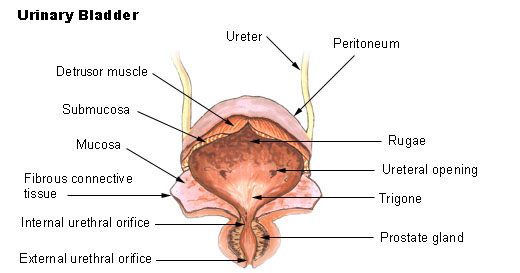 Bladder
 -
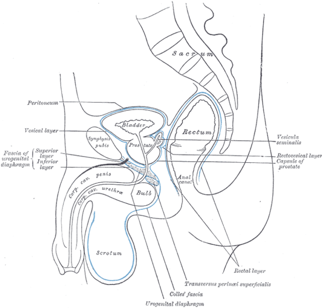 Median sagittal section of pelvis, showing arrangement of fasciæ.
 -
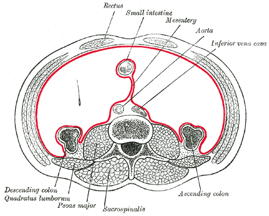 Horizontal disposition of the peritoneum in the lower part of the abdomen.
 -
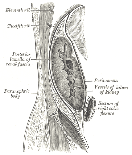 Sagittal section through posterior abdominal wall, showing the relations of the capsule of the kidney.
 -
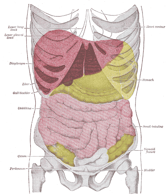 Topography of thoracic and abdominal viscera.


References
- Tortora, Gerard J., Anagnostakos, Reginald Merryweather, Nicholas P. (1984) Principles of Anatomy and Physiology, Harper & Row Publishers, New York ISBN 0-06-046656-1
External links
Template:Peritoneum
Template:Gastroenterology
cs:Pobřišnice
de:Peritoneum
eo:Peritoneo
it:Peritoneo
la:Peritoneum
lt:Pilvaplėvė
nl:Buikvlies
sk:Pobrušnica
sl:Potrebušnica
fi:Vatsakalvo
sv:Bukhinna