Budd-Chiari syndrome differential diagnosis: Difference between revisions
Mazia Fatima (talk | contribs) |
Mazia Fatima (talk | contribs) |
||
| Line 7: | Line 7: | ||
==Overview== | ==Overview== | ||
==Differential diagnosis== | ==Differential diagnosis== | ||
Budd-Chiari syndrome can be differentiated from other causes of hepatic failure and | Budd-Chiari syndrome can be differentiated from other causes of hepatic failure like viral hepatitis and liver cirrhosis. The differential diagnosis is as follows: | ||
<span style="font-size:85%">'''Abbreviations:''' | <span style="font-size:85%">'''Abbreviations:''' | ||
| Line 396: | Line 396: | ||
| style="padding: 5px 5px; background: #F5F5F5;" align="center" |RUQ | | style="padding: 5px 5px; background: #F5F5F5;" align="center" |RUQ | ||
| style="padding: 5px 5px; background: #F5F5F5;" align="center" |Dull / aching | | style="padding: 5px 5px; background: #F5F5F5;" align="center" |Dull / aching | ||
| style="padding: 5px 5px; background: #F5F5F5;" align="center" | + in cirrhotic patients | | style="padding: 5px 5px; background: #F5F5F5;" align="center" | + in [[cirrhotic]] patients | ||
| style="padding: 5px 5px; background: #F5F5F5;" align="center" |may be in cardicmyopathy | | style="padding: 5px 5px; background: #F5F5F5;" align="center" |may be in [[cardicmyopathy]] | ||
| style="padding: 5px 5px; background: #F5F5F5;" align="center" |− | | style="padding: 5px 5px; background: #F5F5F5;" align="center" |− | ||
| style="padding: 5px 5px; background: #F5F5F5;" align="center" |− | | style="padding: 5px 5px; background: #F5F5F5;" align="center" |− | ||
| style="padding: 5px 5px; background: #F5F5F5;" align="center" |>60% TS<br> >240 μg/L SF <br>Raised LFT <br>Hyperglycemia | | style="padding: 5px 5px; background: #F5F5F5;" align="center" |>60% TS<br> >240 μg/L SF <br>Raised LFT <br>[[Hyperglycemia]] | ||
| style="padding: 5px 5px; background: #F5F5F5;" align="center" |Ultrasound shows evidence of cirrhosis | | style="padding: 5px 5px; background: #F5F5F5;" align="center" |Ultrasound shows evidence of [[cirrhosis]] | ||
| style="padding: 5px 5px; background: #F5F5F5;" align="center" |Extra intestinal findings: | | style="padding: 5px 5px; background: #F5F5F5;" align="center" |Extra intestinal findings: | ||
* hyperpigmentation | * hyperpigmentation | ||
* Diabetes mellitus | * [[Diabetes mellitus]] | ||
* Arthralgia | * Arthralgia | ||
* Impotence in males | * [[Impotence]] in males | ||
* Cardiomyopathy | * [[Cardiomyopathy]] | ||
* Atherosclerosis | * [[Atherosclerosis]] | ||
* Hypopituitarism | * [[Hypopituitarism]] | ||
* Hypothyroidism | * [[Hypothyroidism]] | ||
* Extrahepatic cancer | * Extrahepatic [[cancer]] | ||
* Prone to specific infections | * Prone to specific [[infections]] | ||
|- | |- | ||
| style="padding: 5px 5px; background: #DCDCDC;" align="center" |[[Cirrhosis|Cirrhosis]] | | style="padding: 5px 5px; background: #DCDCDC;" align="center" |[[Cirrhosis|Cirrhosis]] | ||
| Line 427: | Line 427: | ||
| style="padding: 5px 5px; background: #F5F5F5;" align="left" | | | style="padding: 5px 5px; background: #F5F5F5;" align="left" | | ||
|- | |- | ||
! style="padding: 5px 5px; background: #DCDCDC;" align="center" | Peritoneal causes | ! style="padding: 5px 5px; background: #DCDCDC;" align="center" | [[Peritoneal]] causes | ||
| colspan="1" rowspan="1" style="padding: 5px 5px; background: #DCDCDC;" align="center" |[[Spontaneous bacterial peritonitis]] | | colspan="1" rowspan="1" style="padding: 5px 5px; background: #DCDCDC;" align="center" |[[Spontaneous bacterial peritonitis]] | ||
| style="padding: 5px 5px; background: #F5F5F5;" align="center" | + | | style="padding: 5px 5px; background: #F5F5F5;" align="center" | + | ||
| Line 441: | Line 441: | ||
* Culture: Positive for single organism | * Culture: Positive for single organism | ||
| style="padding: 5px 5px; background: #F5F5F5;" align="left" |Ultrasound for evaluation of liver cirrhosis | | style="padding: 5px 5px; background: #F5F5F5;" align="left" |Ultrasound for evaluation of liver [[cirrhosis]] | ||
! style="padding: 5px 5px; background: #F5F5F5;" align="left" | | ! style="padding: 5px 5px; background: #F5F5F5;" align="left" | | ||
|- | |- | ||
| Line 481: | Line 481: | ||
| style="padding: 5px 5px; background: #F5F5F5;" align="left" |N | | style="padding: 5px 5px; background: #F5F5F5;" align="left" |N | ||
| style="padding: 5px 5px; background: #F5F5F5;" align="left" |Increased [[bilirubin]] and [[alkaline phosphatase]] | | style="padding: 5px 5px; background: #F5F5F5;" align="left" |Increased [[bilirubin]] and [[alkaline phosphatase]] | ||
| style="padding: 5px 5px; background: #F5F5F5;" align="left" |Ultrasound | | style="padding: 5px 5px; background: #F5F5F5;" align="left" |[[Ultrasound]] | ||
| style="padding: 5px 5px; background: #F5F5F5;" align="left" |[[Nausea and vomiting|Nausea & vomiting]] | | style="padding: 5px 5px; background: #F5F5F5;" align="left" |[[Nausea and vomiting|Nausea & vomiting]] | ||
|- | |- | ||
| Line 494: | Line 494: | ||
| style="padding: 5px 5px; background: #F5F5F5;" align="left" |N | | style="padding: 5px 5px; background: #F5F5F5;" align="left" |N | ||
| style="padding: 5px 5px; background: #F5F5F5;" align="left" |[[Hematuria]] | | style="padding: 5px 5px; background: #F5F5F5;" align="left" |[[Hematuria]] | ||
| style="padding: 5px 5px; background: #F5F5F5;" align="left" |CT scan and ultrasound | | style="padding: 5px 5px; background: #F5F5F5;" align="left" |[[CT scan]] and [[ultrasound]] | ||
| style="padding: 5px 5px; background: #F5F5F5;" align="left" |Colicky [[abdominal pain]] associated with [[Nausea and vomiting|nausea & vomiting]] | | style="padding: 5px 5px; background: #F5F5F5;" align="left" |Colicky [[abdominal pain]] associated with [[Nausea and vomiting|nausea & vomiting]] | ||
|- | |- | ||
| Line 580: | Line 580: | ||
| style="padding: 5px 5px; background: #F5F5F5;" align="left" |[[Vaginal discharge]] | | style="padding: 5px 5px; background: #F5F5F5;" align="left" |[[Vaginal discharge]] | ||
|- | |- | ||
| style="padding: 5px 5px; background: #DCDCDC;" align="center" |Cyst rupture | | style="padding: 5px 5px; background: #DCDCDC;" align="center" |[[Cyst]] rupture | ||
| style="padding: 5px 5px; background: #F5F5F5;" align="center" |− | | style="padding: 5px 5px; background: #F5F5F5;" align="center" |− | ||
| style="padding: 5px 5px; background: #F5F5F5;" align="center" |− | | style="padding: 5px 5px; background: #F5F5F5;" align="center" |− | ||
| Line 590: | Line 590: | ||
| style="padding: 5px 5px; background: #F5F5F5;" align="left" |N | | style="padding: 5px 5px; background: #F5F5F5;" align="left" |N | ||
| style="padding: 5px 5px; background: #F5F5F5;" align="left" |Increased [[ESR]] and [[CRP]] | | style="padding: 5px 5px; background: #F5F5F5;" align="left" |Increased [[ESR]] and [[CRP]] | ||
| style="padding: 5px 5px; background: #F5F5F5;" align="left" |Ultrasound | | style="padding: 5px 5px; background: #F5F5F5;" align="left" |[[Ultrasound]] | ||
| style="padding: 5px 5px; background: #F5F5F5;" align="left" |Sudden onset sever pain with [[nausea and vomiting]] | | style="padding: 5px 5px; background: #F5F5F5;" align="left" |Sudden onset sever pain with [[nausea and vomiting]] | ||
|- | |- | ||
! style="padding: 5px 5px; background: #DCDCDC;" align="center" |Pregnancy | ! style="padding: 5px 5px; background: #DCDCDC;" align="center" |[[Pregnancy]] | ||
| style="padding: 5px 5px; background: #DCDCDC;" align="center" |Ruptured [[ectopic pregnancy]] | | style="padding: 5px 5px; background: #DCDCDC;" align="center" |Ruptured [[ectopic pregnancy]] | ||
| style="padding: 5px 5px; background: #F5F5F5;" align="center" |− | | style="padding: 5px 5px; background: #F5F5F5;" align="center" |− | ||
| Line 604: | Line 604: | ||
| style="padding: 5px 5px; background: #F5F5F5;" align="left" |N | | style="padding: 5px 5px; background: #F5F5F5;" align="left" |N | ||
| style="padding: 5px 5px; background: #F5F5F5;" align="left" |Positive [[pregnancy test]] | | style="padding: 5px 5px; background: #F5F5F5;" align="left" |Positive [[pregnancy test]] | ||
| style="padding: 5px 5px; background: #F5F5F5;" align="left" |Ultrasound | | style="padding: 5px 5px; background: #F5F5F5;" align="left" |[[Ultrasound]] | ||
| style="padding: 5px 5px; background: #F5F5F5;" align="left" |History of missed period and [[vaginal bleeding]] | | style="padding: 5px 5px; background: #F5F5F5;" align="left" |History of missed period and [[vaginal bleeding]] | ||
|- | |- | ||
Revision as of 22:12, 14 November 2017
|
Budd-Chiari syndrome Microchapters |
|
Diagnosis |
|---|
|
Treatment |
|
Case Studies |
|
Budd-Chiari syndrome differential diagnosis On the Web |
|
American Roentgen Ray Society Images of Budd-Chiari syndrome differential diagnosis |
|
Risk calculators and risk factors for Budd-Chiari syndrome differential diagnosis |
Editor-In-Chief: C. Michael Gibson, M.S., M.D. [1]; Associate Editor(s)-in-Chief: Mazia Fatima, MBBS [2] Amandeep Singh M.D.[3]
Please help WikiDoc by adding content here. It's easy! Click here to learn about editing.
Overview
Differential diagnosis
Budd-Chiari syndrome can be differentiated from other causes of hepatic failure like viral hepatitis and liver cirrhosis. The differential diagnosis is as follows:
Abbreviations:
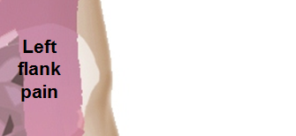
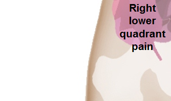
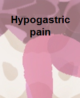
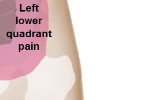
RUQ= Right upper quadrant of the abdomen, LUQ= Left upper quadrant, LLQ= Left lower quadrant, RLQ= Right lower quadrant, LFT= Liver function test, SIRS= Systemic inflammatory response syndrome, ERCP= Endoscopic retrograde cholangiopancreatography, IV= Intravenous, N= Normal, AMA= Anti mitochondrial antibodies, LDH= Lactate dehydrogenase, GI= Gastrointestinal, CXR= Chest X ray, IgA= Immunoglobulin A, IgG= Immunoglobulin G, IgM= Immunoglobulin M, CT= Computed tomography, PMN= Polymorphonuclear cells, ESR= Erythrocyte sedimentation rate, CRP= C-reactive protein
| |||||||||||||||||||||||||||||||||||||||||||||||||||||||||||||||||||||||||||||||||||||||||||||||||||||||||||||||||||||||||||||||||||||||||||||||||||||||||||||||||||||||||||||||||||||||||||||||||||||||||||||||||||||||||||||||||||||||||||||||||||||||||||||||||||||||||||||||||||||||||||||||||||||||||||||||||||||||||||||||||||||||||||||||||||||||||||||||||||||||||||||||||||||||||||||||||||||||||||||||||||||||||||||||||||||||||||||||||||||||||||||||||||||||||||||||||||||||||||||||||||||||||||||||||||||||||||||||||||||||||||||||||||||
 |
 |
|
 |
 |
|
 |
 |
|