Ulcerative colitis physical examination
|
Ulcerative colitis Microchapters |
|
Diagnosis |
|---|
|
Treatment |
|
Case Studies |
|
Ulcerative colitis physical examination On the Web |
|
American Roentgen Ray Society Images of Ulcerative colitis physical examination |
|
Risk calculators and risk factors for Ulcerative colitis physical examination |
Editor-In-Chief: C. Michael Gibson, M.S., M.D. [1]; Associate Editor(s)-in-Chief: Usama Talib, BSc, MD [2]
Overview
Ulcerative colitis shows intestinal and extra intestinal findings on physical examination. These include abdominal tenderness, fever, pallor, inflammation of the iris and uvea, skin rash, inflammation of the joints, aphthous ulcers and clubbing of the fingers.[1][2]
Physical Examination
As ulcerative colitis is a systemic disease, patients may present with symptoms and complications outside the colon. These include the following:[1][3][4][5][6]
General appearance
- Normal appearance in mild disease
- Pallor may be noticed
Vital signs
Gastrointestinal
- Normal in mild disease
- Abdominal tenderness when palpated in severe disease
- Blood seen on rectal exam
Eyes
The ophthalmic physical exam findings may include:
- Iritis or uveitis, which is inflammation of the iris
- Episcleritis
Musculoskeletal
- Seronegative arthritis, which can be a large-joint oligoarthritis (affecting one or two joints), or may affect many small joints of the hands and feet
- Ankylosing spondylitis, arthritis of the spine
- Sacroiliitis, arthritis of the lower spine
Skin
- Erythema nodosum, which is a panniculitis, or inflammation of subcutaneous tissue involving the lower extremities
- Pyoderma gangrenosum, which is a painful ulcerating lesion involving the skin
Other
- Deep venous thrombosis and pulmonary embolism
- Autoimmune hemolytic anemia
- Clubbing, a deformity of the ends of the fingers
- Primary sclerosing cholangitis, or inflammation of the bile ducts
- Aphthous ulcers of the mouth
-
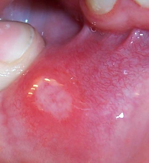 By Photographer: User:TheBlunderbuss - Own work. Published under GFDL in English Wiki, CC BY-SA 3.0, https://commons.wikimedia.org/w/index.php?curid=591677

References
- ↑ 1.0 1.1 Hanauer SB (1996). "Inflammatory bowel disease". N Engl J Med. 334 (13): 841–8. doi:10.1056/NEJM199603283341307. PMID 8596552.
- ↑ Prantera C, Davoli M, Lorenzetti R, Pallone F, Marcheggiano A, Iannoni C; et al. (1988). "Clinical and laboratory indicators of extent of ulcerative colitis. Serum C-reactive protein helps the most". J Clin Gastroenterol. 10 (1): 41–5. PMID 3356884.
- ↑ D'Haens G, Geboes K, Peeters M, Baert F, Ectors N, Rutgeerts P (1997). "Patchy cecal inflammation associated with distal ulcerative colitis: a prospective endoscopic study". Am J Gastroenterol. 92 (8): 1275–9. PMID 9260788.
- ↑ Kim B, Barnett JL, Kleer CG, Appelman HD (1999). "Endoscopic and histological patchiness in treated ulcerative colitis". Am J Gastroenterol. 94 (11): 3258–62. doi:10.1111/j.1572-0241.1999.01533.x. PMID 10566726.
- ↑ Silverberg MS, Satsangi J, Ahmad T, Arnott ID, Bernstein CN, Brant SR; et al. (2005). "Toward an integrated clinical, molecular and serological classification of inflammatory bowel disease: report of a Working Party of the 2005 Montreal World Congress of Gastroenterology". Can J Gastroenterol. 19 Suppl A: 5A–36A. PMID 16151544.
- ↑ Guan Q, Zhang J (2017). "Recent Advances: The Imbalance of Cytokines in the Pathogenesis of Inflammatory Bowel Disease". Mediators Inflamm. 2017: 4810258. doi:10.1155/2017/4810258. PMC 5379128. PMID 28420941.