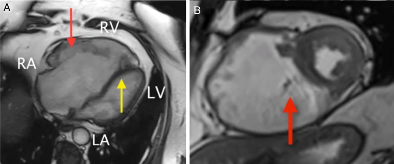
Tricuspid regurgitation cardiac MRI
Jump to navigation
Jump to search
|
Tricuspid Regurgitation Microchapters |
|
Diagnosis |
|---|
|
Treatment |
|
Case Studies |
|
Tricuspid regurgitation cardiac MRI On the Web |
|
American Roentgen Ray Society Images of Tricuspid regurgitation cardiac MRI |
|
Risk calculators and risk factors for Tricuspid regurgitation cardiac MRI |
Editor-In-Chief: C. Michael Gibson, M.S., M.D. [1]; Associate Editor(s)-in-Chief: Vamsikrishna Gunnam M.B.B.S [2] Rim Halaby, M.D. [3]
Overview
Cardiac magnetic resonance imaging (CMR) may be beneficial when echocardiography findings are inconclusive, particularly before tricuspid valve surgery.[1]
Cardiac MRI
- Cardiac MRI is useful in evaluating the structure and function of the right atrium and right ventricle as well as the severity of the tricuspid regurgitation, especially when echocardiography is inconclusive.
- Findings on MRI suggestive of tricuspid regurgitation include:[2][3][4][5][6][7]
- Cardiac magnetic resonance imaging (CMR) helps in giving the quantitative assessment of tricuspid regurgitant volume (TRV)[8]
- Severe tricuspid regurgitation if TMR is ≥30 ml, or in a separate analysis, ≥40 ml
- Moderate or mild tricuspid regurgitation if TMR is < the above threshold.
- Cardiac magnetic resonance imaging (CMR) helps in estimation of regurgitant fraction
- Cardiac magnetic resonance imaging (CMR) helps in estimation of RV volumes and ejection fraction
- Cardiac magnetic resonance imaging (CMR) helps in evaluation of associated left ventricle and mitral disease
- Cardiac magnetic resonance imaging (CMR) helps in identification of TR jet area
- Cardiac magnetic resonance imaging (CMR) helps in giving the quantitative assessment of tricuspid regurgitant volume (TRV)[8]
2014 AHA/ACC Guideline for the Management of Patients With Valvular Heart Disease: Executive Summary[1]
| Class IIb |
| "1. CMR or real-time 3-dimensional echocardiography may be considered for assessment of RV systolic function and systolic and diastolic volumes in patients with severe TR (stages C and D) and suboptimal 2-dimensional echocardiograms. (Level of Evidence: C)" |


References
- ↑ Jump up to: 1.0 1.1 Nishimura RA, Otto CM, Bonow RO, Carabello BA, Erwin JP, Guyton RA; et al. (2014). "2014 AHA/ACC Guideline for the Management of Patients With Valvular Heart Disease: executive summary: a report of the American College of Cardiology/American Heart Association Task Force on Practice Guidelines". Circulation. 129 (23): 2440–92. doi:10.1161/CIR.0000000000000029. PMID 24589852.
- ↑ Zoghbi, William A.; Adams, David; Bonow, Robert O.; Enriquez-Sarano, Maurice; Foster, Elyse; Grayburn, Paul A.; Hahn, Rebecca T.; Han, Yuchi; Hung, Judy; Lang, Roberto M.; Little, Stephen H.; Shah, Dipan J.; Shernan, Stanton; Thavendiranathan, Paaladinesh; Thomas, James D.; Weissman, Neil J. (2017). "Recommendations for Noninvasive Evaluation of Native Valvular Regurgitation". Journal of the American Society of Echocardiography. 30 (4): 303–371. doi:10.1016/j.echo.2017.01.007. ISSN 0894-7317.
- ↑ Driessen MMP, Schings MA, Sieswerda GT, Doevendans PA, Hulzebos EH, Post MC; et al. (2018). "Tricuspid flow and regurgitation in congenital heart disease and pulmonary hypertension: comparison of 4D flow cardiovascular magnetic resonance and echocardiography". J Cardiovasc Magn Reson. 20 (1): 5. doi:10.1186/s12968-017-0426-7. PMC 5767973. PMID 29332606.
- ↑ Reddy ST, Shah M, Doyle M, Thompson DV, Williams RB, Yamrozik J; et al. (2013). "Evaluation of cardiac valvular regurgitant lesions by cardiac MRI sequences: comparison of a four-valve semi-quantitative versus quantitative approach". J Heart Valve Dis. 22 (4): 491–9. PMID 24224411.
- ↑ Mathew RC, Löffler AI, Salerno M (2018). "Role of Cardiac Magnetic Resonance Imaging in Valvular Heart Disease: Diagnosis, Assessment, and Management". Curr Cardiol Rep. 20 (11): 119. doi:10.1007/s11886-018-1057-9. PMC 6415765. PMID 30259253.
- ↑ Zhan, Yang; Debs, Dany; Khan, Mohammad; Nguyen, Duc; Graviss, Edward; Shah, Dipan J. (2019). "CLINICAL OUTCOMES OF SECONDARY TRICUSPID REGURGITATION QUANTIFIED USING CARDIOVASCULAR MAGNETIC RESONANCE". Journal of the American College of Cardiology. 73 (9): 3074. doi:10.1016/S0735-1097(19)33680-0. ISSN 0735-1097.
- ↑ Zhan, Yang; Senapati, Alpana; Vejpongsa, Pimprapa; Xu, Jiaqiong; Shah, Dipan J.; Nagueh, Sherif F. (2020). "Comparison of Echocardiographic Assessment of Tricuspid Regurgitation Against Cardiovascular Magnetic Resonance". JACC: Cardiovascular Imaging. doi:10.1016/j.jcmg.2020.01.008. ISSN 1936-878X.
- ↑ Medvedofsky D, León Jiménez J, Addetia K, Singh A, Lang RM, Mor-Avi V; et al. (2017). "Multi-parametric quantification of tricuspid regurgitation using cardiovascular magnetic resonance: A comparison to echocardiography". Eur J Radiol. 86: 213–220. doi:10.1016/j.ejrad.2016.11.025. PMC 5372350. PMID 28027750.
- ↑ "Gross right heart dilatation secondary to Ebstein's anomaly".
- ↑ "Severe Pulmonary Arterial Hypertension: Comprehensive Evaluation by Magnetic Resonance Imaging".