Erythema nodosum
|
WikiDoc Resources for Erythema nodosum |
|
Articles |
|---|
|
Most recent articles on Erythema nodosum Most cited articles on Erythema nodosum |
|
Media |
|
Powerpoint slides on Erythema nodosum |
|
Evidence Based Medicine |
|
Clinical Trials |
|
Ongoing Trials on Erythema nodosum at Clinical Trials.gov Trial results on Erythema nodosum Clinical Trials on Erythema nodosum at Google
|
|
Guidelines / Policies / Govt |
|
US National Guidelines Clearinghouse on Erythema nodosum NICE Guidance on Erythema nodosum
|
|
Books |
|
News |
|
Commentary |
|
Definitions |
|
Patient Resources / Community |
|
Patient resources on Erythema nodosum Discussion groups on Erythema nodosum Patient Handouts on Erythema nodosum Directions to Hospitals Treating Erythema nodosum Risk calculators and risk factors for Erythema nodosum
|
|
Healthcare Provider Resources |
|
Causes & Risk Factors for Erythema nodosum |
|
Continuing Medical Education (CME) |
|
International |
|
|
|
Business |
|
Experimental / Informatics |
Editor-In-Chief: C. Michael Gibson, M.S., M.D. [1]; Associate Editor(s)-in-Chief: Kiran Singh, M.D. [2]
Overview
Erythema nodosum (EN) (red nodules) is an inflammation of the fat cells under the skin (panniculitis). It causes tender, red nodules that are usually seen on both shins. EN is an immunologic response to a variety of different causes.
Historical Perspective
- Disease name] was first discovered by [scientist name], a [nationality + occupation], in [year] during/following [event].
- In [year], [gene] mutations were first identified in the pathogenesis of [disease name].
- In [year], the first [discovery] was developed by [scientist] to treat/diagnose [disease name].
Classification
- [Disease name] may be classified according to [classification method] into [number] subtypes/groups:
- [group1]
- [group2]
- [group3]
- Other variants of [disease name] include [disease subtype 1], [disease subtype 2], and [disease subtype 3].
Pathophysiology
- It is considered as a hypersensitivity reaction to various antigens leading to deposition of immune complexes in subcutaneous fat venules. Evidence of immune complexes and complement in the early lesions is affirmative of the above hypothesis.
- Alternative hypothesis involving type 4 hypersensitive reaction, expression of various adhesion and inflammatory markers like P-selectin, E-selectin, IL-6, IL-8 has also been proposed in the pathophysiology of erythema nodosum. (Springer)
Causes
- In about 30-50% of cases, the cause of EN is unknown. Idiopathic is the most common cause.[1]
- EN may be associated with a wide variety of diseases including the following
- Infections (e.g., Tuberculosis, Streptococcal, Mycoplasma pneumoniae, and Epstein-Barr virus)
- Sarcoidosis
- Inflammatory bowel disease
- Autoimmune disorders (e.g., Behçet's disease)
- Pregnancy
- Medications (sulfonamides, clomiphene, oral contraceptives (Desogestrel and Ethinyl Estradiol), bromides, certolizumab pegol, dapsone), and cancer.[1][2][3][4]
Differentiating Erythema nodosum from Other Diseases
Erythema nodosum is indicative of an underlying infectious disease but a cause is not found in about half the cases.[5][6]
- Behcet's Syndrome
- Brucellosis
- Campylobacter
- Cat-Scratch Fever
- Chlamydia
- Coccidioidomycosisimportant in the south-west USA
- Corynebacterium diphtheria
- Crohn's Disease
- Dermatophytosis
- Drugs such as sulphonylureas, gold and oral contraceptives
- Francisella tularensis
- Hepatitis
- Herpes simplex
- Histoplasmosis
- Hodgkin's Lymphoma can precede the diagnosis
- Hookworm infection
- Infectious mononucleosis
- Leprosymay closely resemble erythema nodosum but the histological findings are different
- Leptospirosis
- Leukemia
- Milker's nodule
- Mycobacterium
- Mycoplasma pneumoniae
- Neisseria Meningitidis
- North American Blastomycosis
- Postradiated pelvic cancer
- Pregnancy usually in the second trimester. Often recurs during future pregnancies and may occur with oral contraceptive uses
- Radiation therapy
- Reiter's Disease
- Salmonella
- Sarcoidosis
- Sporotrichosis
- Streptococcal infections are one of the most common causes
- Toxoplasmosis
- Tuberculosis
- Ulcerative colitis
- Yersinia
Epidemiology and Demographics
Erythema nodosum is the most common form of panniculitis (inflammation of the subcutaneous fat). The prevalence is 24 cases per 100,000 per year. The peak incidence of EN occurs between 20-30 years of age. Women are 3-6 times more commonly affected than men[7].
Age: 20 to 30
Gender: Female > Male ( 6:1)
Race: African americans ( AAFP)
Risk Factors
Natural History, Complications and Prognosis
- The majority patients with Erythema Nodosum will recover completely in few weeks.
- Common complications of [disease name] include [complication 1], [complication 2], and [complication 3].
- Prognosis is generally excellent. .
Diagnosis
Diagnostic Criteria
Diagnosis is clinical. A deep punch biopsy or an incisional biopsy may be performed in cases where the diagnosis is unclear. Microscopic examination will reveal a septal panniculitis with acute and chronic inflammation in the fat and around blood vessels[7].
Once EN is diagnosed, additional evaluation needs to be performed to determine the underlying cause. A complete blood count, erythrocyte sedimentation rate (ESR), antistreptolysin-O (ASO) titer, urinalysis, throat culture, intradermal tuberculin test, and chest x-ray is part of the initial examination.[8]
The ESR is initially very high, and falls as the nodules fade. The ASO titer is high in cases associated with a streptococcal throat infection. A chest X-ray should be performed to rule out pulmonary diseases. Hilar lymphadenopathy may be due to tuberculosis, sarcoidosis, or Löfgren syndrome (a form of acute sarcoidosis with erythema nodosum , parotid swelling and bilateral hilar adenopathy, often accompanied by joint symptoms).
Symptoms
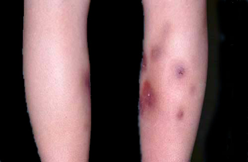
Erythema nodosum occurs 3-6 weeks after an event, either internal or external to the body, that initiates a hypersensitivity reaction in subcutaneous fat [9][10]. EN is frequently associated with fever, malaise, arthralgia, and joint pain and inflammation. It presents as tender red nodules on the shins that are smooth and shiny. The nodules may occur anywhere there is fat under the skin, including the thighs, arms, trunk, face, and neck [11][12]. The nodules are 1-5 cm in diameter, and individual nodules may coalesce to form large areas of hardened skin.
As the nodules age, they become bluish purple, brownish, yellowish, and finally green, similar to the color changes that occur in a resolving bruise. The nodules usually subside over a period of 2–6 weeks without ulceration or scarring[7].
Dermatophytids are similar skin lesions that result from a fungus infection such as ringworm in another area of the body.
Physical Examination
Skin
-
Erythema Nodosum involving the lower extremity
-
![Erythema Nodosum[13]](/images/d/d5/Erythema_nodosum_%28IBD%2C_Coccidoides_imitis%2C_TB%2C_Sarcoid%29.jpg)
Erythema Nodosum[13]

![Erythema Nodosum[13]](/index.php/File:Erythema_nodosum_(IBD,_Coccidoides_imitis,_TB,_Sarcoid).jpg)
Extremities
-
![.:Erythema nodosum Adapted from Dermatology Atlas.[14]](/images/d/d0/Erythema_nodosum01.jpg)
.:Erythema nodosum Adapted from Dermatology Atlas.[14]
-
![.:Erythema nodosum Adapted from Dermatology Atlas.[14]](/images/3/37/Erythema_nodosum02.jpg)
.:Erythema nodosum Adapted from Dermatology Atlas.[14]
-
![.:Erythema nodosum Adapted from Dermatology Atlas.[14]](/images/8/84/Erythema_nodosum03.jpg)
.:Erythema nodosum Adapted from Dermatology Atlas.[14]
-
![.:Erythema nodosum Adapted from Dermatology Atlas.[14]](/images/c/c1/Erythema_nodosum04.jpg)
.:Erythema nodosum Adapted from Dermatology Atlas.[14]
-
![.:Erythema nodosum Adapted from Dermatology Atlas.[14]](/images/d/dc/Erythema_nodosum05.jpg)
.:Erythema nodosum Adapted from Dermatology Atlas.[14]
-
![.:Erythema nodosum Adapted from Dermatology Atlas.[14]](/images/7/7e/Erythema_nodosum06.jpg)
.:Erythema nodosum Adapted from Dermatology Atlas.[14]
![.:Erythema nodosum Adapted from Dermatology Atlas.[14]](/index.php/File:Erythema_nodosum01.jpg)
![.:Erythema nodosum Adapted from Dermatology Atlas.[14]](/index.php/File:Erythema_nodosum02.jpg)
![.:Erythema nodosum Adapted from Dermatology Atlas.[14]](/index.php/File:Erythema_nodosum03.jpg)
![.:Erythema nodosum Adapted from Dermatology Atlas.[14]](/index.php/File:Erythema_nodosum05.jpg)
![.:Erythema nodosum Adapted from Dermatology Atlas.[14]](/index.php/File:Erythema_nodosum06.jpg)
- Lesions begin as red, tender nodules. The borders are poorly defined and they are 2 to 6 cms in diameter
- In the first week the lesions become tense, hard, and painful. In the second week, they may become fluctuant, rather like an abscess, but they do not suppurate or ulcerate. Individual lesions last around 2 weeks, but occasionally, new lesions continue to appear for 3 to 6 weeks.
- Initially, in the first week the lesions are bright red but in the second week they assume a more blue or purple hue.
- The lesions may eventually even turn yellow like a bruise which is resolving before they disappear in a several weeks.
- Usually the rash appears on the extremities, but most frequently they occur on the anterior aspect of the lower leg.
- When the underlying cause is an infection, the lesions usually heal in 6 to 8 weeks.
- If the cause is idiopathic, 30% of cases last 6 months.
Laboratory Findings
Other Imaging Findings
Other Diagnostic Studies
Treatment
Medical Therapy
Treatment should focus on the underlying cause. Symptoms can be treated with bedrest, leg elevation, compressive bandages, wet dressings, and nonsteroidal anti-inflammatory agents (NSAIDs).[7] NSAIDS are usually more effective at the onset of EN versus with chronic disease. Steroids can be of benefit but may be contraindicated if infection is present. In erythema nodosum associated with leprosy, thalidomide may be helpful.
Potassium iodide can be used for persistent lesions whose cause remains unknown. Corticosteroids and colchicine can be used in severe refractory cases (Yurdakul et al, 2001).[15][16][17]
Surgery
Prevention
References
- ↑ 1.0 1.1 Mert A, Ozaras R, Tabak F, Pekmezci S, Demirkesen C, Ozturk R (2004). "Erythema nodosum: an experience of 10 years". Scand J Infect Dis. 36 (6–7): 424–7. PMID 15307561.
- ↑ Anan T, Imamura T, Yokoyama S, Fujiwara S (2004). "Erythema nodosum and granulomatous lesions preceding acute myelomonocytic leukemia". J Dermatol. 31 (9): 741–7. PMID 15628321.
- ↑ Bohn S, Buchner S, Itin P (1997). "[Erythema nodosum: 112 cases. Epidemiology, clinical aspects and histopathology]". Schweiz Med Wochenschr. 127 (27–28): 1168–76. PMID 9324739.
- ↑ Lin JT, Chen PM, Huang DF, Kwang WK, Lo K, Wang WS (2004). "Erythema nodosum associated with carcinoid tumour". Clin Exp Dermatol. 29 (4): 426–7. PMID 15245549.
- ↑ "StatPearls". 2021. PMID 29262192.
- ↑ Leung, Alexander K. C.; Leong, Kin Fon; Lam, Joseph M. (2018). "Erythema nodosum". World Journal of Pediatrics. 14 (6): 548–554. doi:10.1007/s12519-018-0191-1. ISSN 1708-8569.
- ↑ 7.0 7.1 7.2 7.3 Wolff K, Johnson RA, Suurmond R (2005). "Section 7: Miscellaneous inflammatory disorders". Fitzpatrick's Color Atlas & Synopsis of Clinical Dermatology (5th ed. ed.). New York, NY: McGraw-Hill Professional. ISBN 0-07-144019-4.
- ↑ Garcia-Porrua C, Gonzalez-Gay MA, Vazquez-Caruncho M, Lopez-Lazaro L, Lueiro M, Fernandez ML, Alvarez-Ferreira J, Pujol RM (2000). "Erythema nodosum: etiologic and predictive factors in a defined population". Arthritis Rheum. 43 (3): 584–92. PMID 10728752.
- ↑ Ryan TJ (1998). "Cutaneous vasculitis". In Burns DA, Breathnach SM. Textbook of Dermatology (6th ed. ed.). London: Blackwell Science Ltd. pp. 2155–225. ISBN 0-632-05064-0.
- ↑ Camilleri MJ, Su WPD (2003). "Panniculitis". In Freedberg IM, Eisen AZ, Wolff K, Austen KF, Goldsmith LA, Katz SI. Fitzpatrick's Dermatology In General Medicine (6th ed. ed.). New York, NY: McGraw-Hill. ISBN 0-07-138076-0.
- ↑ Cribier B, Caille A, Heid E, Grosshans E (1998). "Erythema nodosum and associated diseases. A study of 129 cases". Int J Dermatol. 37 (9): 667–72. PMID 9762816.
- ↑ White WL, Hitchcock MG (1999). "Diagnosis: erythema nodosum or not?". Semin Cutan Med Surg. 18 (1): 47–55. PMID 10188842.
- ↑ http://picasaweb.google.com/mcmumbi/USMLEIIImages
- ↑ 14.0 14.1 14.2 14.3 14.4 14.5 "Dermatology Atlas".
- ↑ Mat C, Yurdakul S, Uysal S, Gogus F, Ozyazgan Y, Uysal O, Fresko I, Yazici H (2005). "A double-blind trial of depot corticosteroids in Behcet's syndrome". Rheumatology (Oxford). PMID 16263779.
- ↑ Wallace SL (1967). "Erythema nodosum treatment with colchicine". JAMA. 202 (11): 1056. PMID 6072607.
- ↑ Yurdakul S, Mat C, Tuzun Y, Ozyazgan Y, Hamuryudan V, Uysal O, Senocak M, Yazici H (2001). "A double-blind trial of colchicine in Behcet's syndrome". Arthritis Rheum. 44 (11): 2686–92. PMID 11710724.
Additional Resource
- Sailer, Christian, Wasner, Susanne. Differential Diagnosis Pocket. Hermosa Beach, CA: Borm Bruckmeir Publishing LLC, 2002:131-132 ISBN 1591032016