Ventricular tachycardia classification: Difference between revisions
| Line 68: | Line 68: | ||
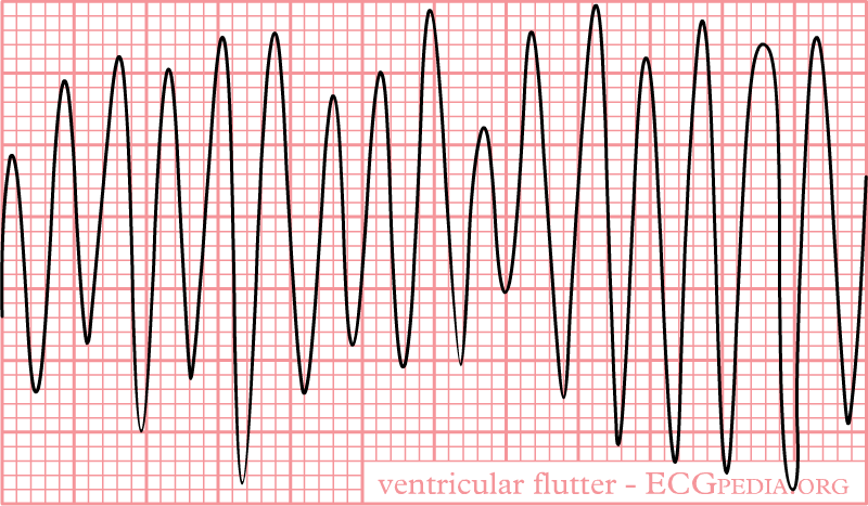
* [[Sinusoidal]] [[monophorphic]] [[QRS]] complexes | * [[Sinusoidal]] [[monophorphic]] [[QRS]] complexes | ||
* [[Without]] any isoelecterical interval between successive [[QRS]] complexes | * [[Without]] any isoelecterical interval between successive [[QRS]] complexes | ||
|[[File:Capture Ven Flu.PNG|center|300px]]<ref> ECG found in https://en.ecgpedia.org/index.php?title=Main_Page </ref> | |[[File:Capture Ven Flu.PNG|center|300px]]<ref> ECG found in https://en.ecgpedia.org/index.php?title=Main_Page </ref> | ||
| | | | ||
Revision as of 07:34, 26 April 2021
|
Ventricular tachycardia Microchapters |
|
Differentiating Ventricular Tachycardia from other Disorders |
|---|
|
Diagnosis |
|
Treatment |
|
Case Studies |
|
Ventricular tachycardia classification On the Web |
|
to Hospitals Treating Ventricular tachycardia classification |
|
Risk calculators and risk factors for Ventricular tachycardia classification |
Editor-In-Chief: C. Michael Gibson, M.S., M.D. [1]; Associate Editor-In-Chief: Cafer Zorkun, M.D., Ph.D. [2]
Overview
Ventricular tachycardia refers to a rhythm with a heart rate in excess of 100 (and in some definitions 120) beats per minute that arises distal to the bundle of His. Ventricular tachycardia can be classified based on three main categories. Firstly, The morphology of the QRS complexes on the ECG (monomorphic ventricular tachycardia vs polymorphic ventricular tachycardia). Secondly, The duration of the episode. Thirdly, The symptoms associated with the episode.
Classification Based Upon Morphology of the QRS Complexes
Classification of ventriculat arrhythmia:
| Term | Definition | Feature | |
|---|---|---|---|
| Ventricular tachycardia | Presence of≥ 3 consecutive premature ventricular complexes with the rate of >100 beats per minute or cycle length< 600 ms |
|
|
| Sustained VT |
|
||
| Nonsustained, or unsustained VT |
|
||
| Monomorphic VT |
|
||
| Polymorphic VT | |||
| Bidirectional VT |
|
||
| Torsades de pointes |
|
 |
|
| Ventricular flutter |
|
 |
|
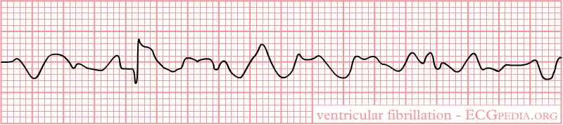
| Ventricular fibrillation |
|
https://nl.ecgpedia.org/images/9/9e/Rhythm_ventricular_fibrillation.png
https://nl.ecgpedia.org/images/thumb/9/9e/Rhythm_ventricular_fibrillation.png/120px-Rhythm_ventricular_fibrillation.png
https://nl.ecgpedia.org/images/f/f6/Rhythm_flutter.png
https://en.ecgpedia.org/index.php?title=Torsade_de_Pointes
Based on the morphology of QRS complex ventricular tachycardia can be classified into the following subtypes:
Monomorphic ventricular tachycardia
- Monomorphic ventricular tachycardia is defined as a QRS complex morphology that is identical from beat-to-beat in all leads of the surface electrocardiogram (ECG).

Polymorphic ventricular tachycardia
- Polymorphic ventricular tachycardia is defined as QRS complexes with beat-to-beat variability in morphology.
- This most commonly appears as a cyclical progressive change in cardiac axis referred to by its french eponym torsade de pointes (literally translating to "twisting of the points").

Classification Based Upon Duration of Episode
Based upon duration of episode ventricular tachycardia can be classified into:
Non-sustained Ventricular Tachycardia or Paroxysmal Ventricular Tachycardia
- Rapid succession of three or more ectopic beats.
- Self terminates within 30 seconds.
Sustained Ventricular Tachycardia
- Lasts more than 30 seconds.
Incessant Ventricular Tachycardia
- The ventricular tachycardia is recurrent and the episodes are interrupted by only a few sinus beats.
Classification Based Upon Symptoms
Based upon symptoms ventricular tachycardia can be classified into:
Pulseless VT
- Pulseless VT is associated with no effective cardiac output, hence, no effective pulse, and is a cause of cardiac arrest.
- In this circumstance it is best treated the same way as ventricular fibrillation (VF) and is recognized as one of the shockable rhythms on the cardiac arrest protocol.
Perfusing Rhythm with VT
- Some VT is associated with reasonable cardiac output and may even be asymptomatic.
- The heart usually tolerates this rhythm poorly in the medium to long term, and patients may certainly deteriorate to pulseless VT or to VF.
References
- ↑ ECG found in https://en.ecgpedia.org/index.php?title=Main_Page
- ↑ ECG found in https://en.ecgpedia.org/index.php?title=Main_Page