Venous insufficiency: Difference between revisions
| Line 137: | Line 137: | ||
If after removal of the below-knee tourniquet the lesser saphenous system fills, then presence of lesser saphenous incompetence is most likely. In following step, the above-knee tourniquet is removed to assess the competence of the [[Hunter's canal]] perforator. At the end, if the superficial venous system remains empty, then the high thigh tourniquet is removed to detect saphenofemoral incompetence. | If after removal of the below-knee tourniquet the lesser saphenous system fills, then presence of lesser saphenous incompetence is most likely. In following step, the above-knee tourniquet is removed to assess the competence of the [[Hunter's canal]] perforator. At the end, if the superficial venous system remains empty, then the high thigh tourniquet is removed to detect saphenofemoral incompetence. | ||
===Trendelenburg test=== | ===Trendelenburg test=== | ||
A '''Trendelenburg test''' determines the competency of the [[Vein valve|valves]] in communicating veins between the [[Superficial vein|superficial]] and [[deep vein]]s of the leg. The leg is raised above heart level until the veins become empty, then the leg is quickly lowered. Superficial veins of the leg normally empty into deep veins, however retrograde filling occurs when valves are incompetent, leading to [[varicose veins]]. | |||
The Trendelenburg test is often confused with [[Trendelenburg's sign]], which is related to conditions affecting the hip and femur. | |||
===Perthes test=== | ===Perthes test=== | ||
Revision as of 06:45, 20 January 2009
| Venous insufficiency | |
 | |
|---|---|
| Venous insufficiency |
Editor-In-Chief: C. Michael Gibson, M.S., M.D. [1]
Please Take Over This Page and Apply to be Editor-In-Chief for this topic: There can be one or more than one Editor-In-Chief. You may also apply to be an Associate Editor-In-Chief of one of the subtopics below. Please mail us [2] to indicate your interest in serving either as an Editor-In-Chief of the entire topic or as an Associate Editor-In-Chief for a subtopic. Please be sure to attach your CV and or biographical sketch.
Overview
Venous insufficiency is a condition in which the veins do not efficiently return blood from the lower limbs back to the heart. Venous insufficiency involves one or more veins.
Normal Anatomy and Function
- The superficial veins lie in the subcutaneous fatty layer of the body just beneath the skin and superficial to the deep fascia enveloping the body musculature. The principal veins in the legs are the great and lesser saphenous veins and their tributaries; in the arms they are the basilic and cephalic veins and their tributaries.
- The deep veins accompany arteries and bear the same name as the arteries they parallel. It is common in the extremities for there to be two or more veins accompanying a small to medium sized artery.
- The perforating veins penetrate the deep fascia and connect the superficial veins to the deep veins. Those along the inner (medial) side of the lower leg play a major role in the pathogenesis of the postphlebitic leg.
- The intramuscular sinusoidal veins are large, very thin walled, valveless veins within skeletal muscle. They connect directly with the deep veins.
-
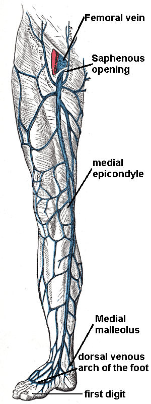
Great saphenous vein
-
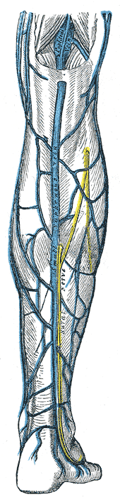
Small saphenous vein and its tributaries.
-
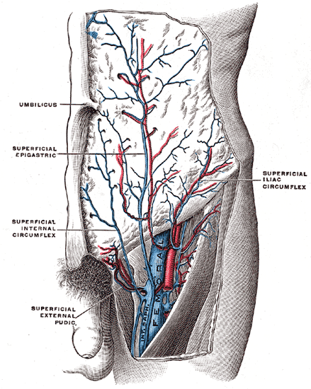
The femoral vein and its tributaries.

Function

Veins serve to return blood from organs to the heart.
In systemic circulation oxygenated blood is pumped by the left ventricle through the arteries to the muscles and organs of the body, where its nutrients and gases are exchanged at capillaries, entering the veins filled with cellular waste and carbon dioxide.
The de-oxygenated blood is taken by veins to the right atrium of the heart, which transfers the blood to the right ventricle, where it is then pumped to the pulmonary arteries and eventually the lungs.
In pulmonary circulation the pulmonary veins return oxygenated blood from the lungs to the left atrium, which empties into the left ventricle, completing the cycle of blood circulation. Normal venous flow is dependent on four factors:
- Dynamic Flow: The heart related flow (dynamics / spontaneous flow). Flow in the arterial system is dependent on the pumping action of the heart and the elasticity and muscular activity of the arteries.
- Phasic Flow: The breathing-related intra-abdominal pressure changes lead to respiratory fluctuation of venous flow with faster flow during expiration due to lower intraabdominal pressure (upward movement of diaphragm) and slower flow during inspiration due to higher intraabdominal pressure (downward movement of diaphragm). This pressure dependent flow pattern is transmitted through the upper leg veins into the major deep veins in the distal lower leg and into the major superficial veins (great and small saphenous veins) in the recumbent patient.
- The muscle pump or the venous pump: The muscle pump mechanism is highly developed in the calf muscles. Large venous sinusoids located in these muscles. As they contract, the force helps to emptying the below veins. Contractions of the calf muscles can produce a sufficient pressure to empty the sinusoids into the deep veins. The deep veins are affected with the similar compressing force due to a strong fascial structure. As a result, with each muscle contraction venous blood is pumped towards to the heart.
- The valves: The valves are prevent retrograde flow. They prevent retrograde flow from heart to veins and from deep veins to superfacial veins.
Pathophysiology

Disturbed venous return from peripheral veins) can have the following causes:
- Impairment of the calf muscle pump
- Obstruction of the deep veins
- Valve incompetence of the epifascial veins
- Valve incompetence of the perforating veins
- Valve incompetence of the subfascial veins
Risk Factors
- Ageing
- Family history
- Female hormones
- Gravitational hydrostatic forces (exacerbated during pregnancy)
- Hydrostatic muscular compartment force
- Sedentary lifestyle
Classification
CEAP Classification: Clinical Presentation, Etiology, Anatomical Localization, Pathophysiological Dysfunction
- According to Clinical Presentation
- Asymptomatic
- Symptomatic
- According to Etiology
- Congenital
- Primary
- Secondary)
- According to Anatomical Localization / Distribution
- Superficial
- Deep
- Perforator
- Alone
- In combination
- According to Pathophysiological Dysfunction
- Reflux
- Obstruction
- Alone
- In combination
Evaluation
- Class 0: No evidence of venous disease.
- Class 1: Telangiectasia (Superficial spider veins), reticular veins, malleolar flare
- Class 2: Simple varicose veins only
- Class 3: Ankle oedema of venous origin (not foot edema)
- Class 4: Skin pigmentation in the gaiter area (lipodermatosclerosis)
- Class 5: Dermatological changes with a healed venous ulcer
- Class 6: Dermatological changes with an open (active) venous ulcer
Diagnosis
Clinical manifestations of venous insufficiency include various conditions such as telangiectasias, varicose veins, and axial incompetence. These are easily treated. In contrast, venous insufficiency may be refractory to treatment as in the severely damaged postthrombotic limb which manifests segmental occlusion in combination with universal venous reflux.
Signs and Symptoms
- Leg discomfort and / or pain: Complains may include dull aching, heaviness, or cramping. Venous claudication may mimic arterial intermittent claudication, though it typically takes longer to subside after stopping exercise.
- Skin pigmentation and discoloration of the skin. Venous insufficiency is characterized by a dark bluish / purple discoloration. Over time, long standing stasis of blood leads to the deposition of hemosiderin, giving the skin a dark, speckled appearance. If the leg is placed in a dependent position, the bluish/purple discoloration may darken dramatically, further suggestive of venous insufficiency. This occurs as a result of gravity working against an already ineffective blood return system. Patients with severe arterial insufficiency, on the other hand, may have relatively pale skin as a result of under perfusion. When their legs are placed in a dependent position, gravity enhances arterial inflow and the skin may become more red as maximally dilated arterioles attempt to bring blood to otherwise starved tissues. In cases of severe ischemia, the affected areas (usually involving the most distal aspect of the foot), can appear whitish or mottled, giving the leg a marbleized appearance. Dead tissue turns black (a.k.a. gangrene). Cellulitis (infection in the skin) will cause the skin to appear bright red. These changes can be difficult to detect in people of color.
- Ulceration: Non-healing ulcers especially around the medial malleolus
- Lipodermatosclerosis: LDS or liposclerosis refers to a thickening in the tissues underneath the skin.
- Varicose eczema: the skin becomes red, wet and scaly.
- Leg swelling
Physical Examination
- Auscultation findings of arteriovenous fistulas may be present
- Increased skin temperature may found
- Thin skin: Sign of poor skin nutrition.
Tourniquet tests
Performing a Tourniquet test is necessary to determine the level of valvular incompetence in the superficial system and to ascertain whether deep venous system involvement is present. Tourniquet test is developed by Brody and later on modified by Trendelenburg.
The patient should wait in a supine position with the limb elevated for at least one minute before starting to perform the Tourniquet test. This maneuver empties the veins by reducing venous congestion in the superficial venous system. Four tourniquets are then placed at the upper thigh, lower thigh, calf, and upper ankle.
Then ask to patient to stand. If the superficial veins of the calf segment fill, perforating vein incompetence is usually present. The tourniquets are then removed from the bottom upward. If removing the ankle tourniquet fills the superficial venous system, presence of perforating vein incompetence is suspected.
If after removal of the below-knee tourniquet the lesser saphenous system fills, then presence of lesser saphenous incompetence is most likely. In following step, the above-knee tourniquet is removed to assess the competence of the Hunter's canal perforator. At the end, if the superficial venous system remains empty, then the high thigh tourniquet is removed to detect saphenofemoral incompetence.
Trendelenburg test
A Trendelenburg test determines the competency of the valves in communicating veins between the superficial and deep veins of the leg. The leg is raised above heart level until the veins become empty, then the leg is quickly lowered. Superficial veins of the leg normally empty into deep veins, however retrograde filling occurs when valves are incompetent, leading to varicose veins.
The Trendelenburg test is often confused with Trendelenburg's sign, which is related to conditions affecting the hip and femur.
Perthes test
The Perthes test is a clinical test for assessing the patency of the deep femoral vein prior to varicose vein surgery. It is named after German surgeon Georg Perthes.
The limb is elevated and an elastic bandage is applied firmly from the toes to the upper 1/3 of the thigh to obliterate the superficial veins only.
With the bandage applied the patient is asked to walk for 5 minutes. If deep system is competent, the blood will go through and back to the heart. If the deep system is incompetent, the patient will feel pain in the leg.
This test is sometimes referred to as the Delbet-Mocquot test, named after French physicians Pierre Delbet and Pierre Mocquot.
Modified Perthes test
The test in done by applying a tourniquet at the level of the saphenofemoral junction to occlude the superficial pathway, and then the patient is asked to move in situ. If the deep veins are occluded, the dilated veins increase in prominence and pain occurs.
This is a more reliable test as it does not depend on patient's pain threshold.
Ultrasonography
Gray scale ultrasonography (Compress ultrasonography)
Color Doppler Ultrasonography
Duplex scanning of the deep and superficial veins can detect obstruction. In addition, the function of valves in each segment of the evaluated veins can be assessed by determining the direction of blood-flow using Doppler ultrasound. Function of the proximal valves is evaluated during Valsalva maneuver in the recumbent patient and Doppler sampling in the common and superficial femoral veins during increased abdominal pressure.
The examination is often done in the upright position, as this is the best way to evaluate valve function.
Landmarks for venous reflux examination:
- Common femoral vein
- Femoral vein
- Upper third part
- Distal third part
- Popliteal vein
- Gastrocnemius veins
- Saphenofemoral junction
- Saphenous vein (above the knee)
- Saphenous vein (below the knee)
- Saphenopopliteal junction
- Mode of termination, short saphenous vein
Therapeutic decision depends on color flow duplex ultrasound evaluation results. Following an examination there are four main levels of venous pathology:
- Superficial venous reflux only
- Deep venous reflux only
- Mixed superficial and deep venous reflux
- Occluded deep venous system
Photoplethysmography (PPG)
Air Plethysmography (APG)
Mercury Strain-gauge Plethysmography
Real-Time Digital Phlebography
Light Reflection Rheography
MRI
MRI provides sagittal, coronal, and cross-sectional views and is able to detect acute occlusion.
Computed Tomography
Phlebography
- Ascending
- Descending
Differential diagnosis of causes of venous insufficiency
Treatment
Medical Management
- Elevation of the leg
- Exercise
- Elastic compression (Compression stockings)
Compression Therapy: External banding to restore saphenous competence
- Graduated compression stockings
- Custom made
- Standard size
- Knee length
- Thigh length
- Compression tights
- Bandages
- Single component
- Multiple components
- Inelastic
- Elastic
- Intermittent pneumatic compression
- Single chamber
- Sequential chambers
- Foot-pump
- Lower leg
- Full leg & trunk
Stent Application
Self-expanding metallic stents are used in venous stenting.
Hemodynamic correction of varicose veins (CHIVA)
Surgery
- Superficial Venous Stripping
- Conventional surgery: Conventional surgery for varicose veins does relieve symptoms and has a role on the prevention of chronic venous ulceration.
- Valve repair techniques
- Techniques with Phlebotomy
- Internal Valvuloplasty
- Venous Segment Transfer
- Vein Valve Transplantation
- Neo Valve
- Allograft Cryopreserved Valve
- Techniques without Phlebotomy
- Wrapping, Banding, Cuffing, and External Stenting
- External Valvuloplasty
- Transmural Valvuloplasty
- Transcommissural Valvuloplasty
- Angioscopy-Assisted External Valve Repair
- Percutaneously placed device
- The Portland valve
Sclerotherapy
- Conventional Sclerotherapy
- Foam Sclerotherapy
Endovenous Ablation (Saphenous Venous Ablation - Closure)
- Endovenous laser ablation (EVLA) is a relatively simple and quick technique which can be performed under a local anesthetic. Endovenous laser techniques employ an 810 nm-diode laser to heat the long or short saphenous vein (or major tributaries), inducing a combination of endothelial damage, focal coagulative necrosis, shrinkage of the vein and thrombotic occlusion.
- Radiofrequency ablation
Complications
Prevention
Examination Findings
-

Venous insufficiency
-

-

Assymetric Leg, Swelling secondary to Deep Venous Thrombosis in Right Leg
-
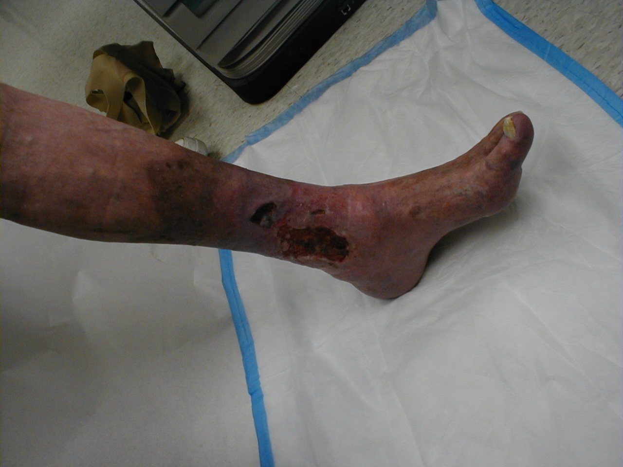
Venous Stasis Ulcer
-
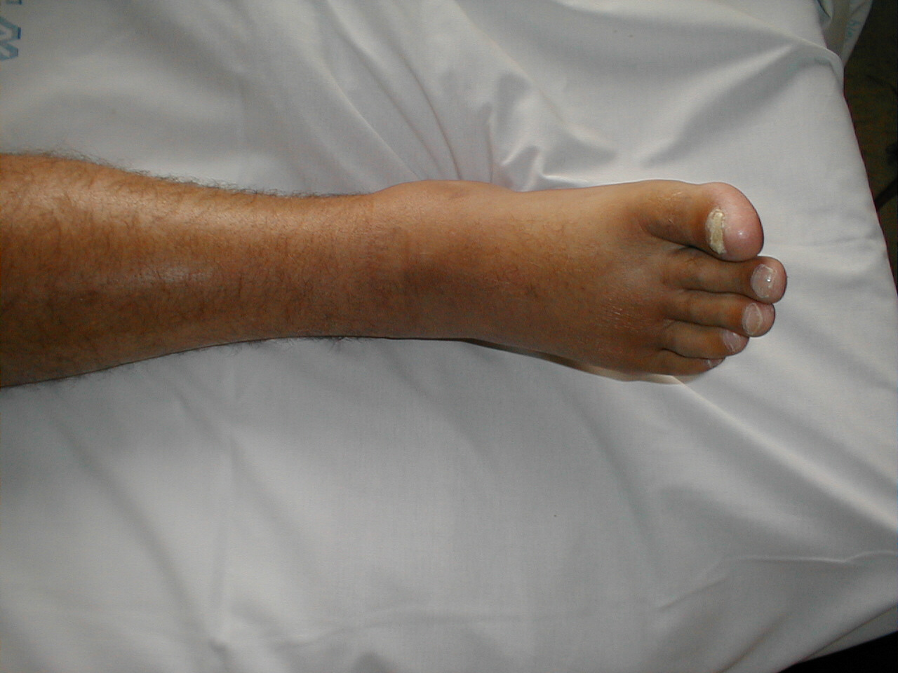
Edema of Right Foot
-

Massive edema


References
Additional Resources
See Also
External Links
Template:Veins Template:VeinsHeadNeck Template:Veins of the upper extremity Template:Veins of the lower extremity Template:SIB