Tuberculosis laboratory findings: Difference between revisions
| Line 11: | Line 11: | ||
The reaction should be measured in millimeters of the induration (palpable, raised, hardened area or swelling). The reader should not measure erythema (redness). The diameter of the indurated area should be measured across the forearm (perpendicular to the long axis). | The reaction should be measured in millimeters of the induration (palpable, raised, hardened area or swelling). The reader should not measure erythema (redness). The diameter of the indurated area should be measured across the forearm (perpendicular to the long axis). | ||
====Classification of Tuberculin Reaction <ref>{{cite web|url= http://www.cdc.gov/tb/publications/factsheets/testing/skintesting.htm|title= CDC Tuberculin Skin Testing| }}</ref>==== | ====Classification of Tuberculin Reaction <ref>{{cite web|url= http://www.cdc.gov/tb/publications/factsheets/testing/skintesting.htm|title= CDC Tuberculin Skin Testing| }}</ref>==== | ||
{| | {|valign=top| | ||
[[File:Mantoux tuberculin skin test.jpg|thumb|200px|right|Image from Public Health Image Library (PHIL)]] | [[File:Mantoux tuberculin skin test.jpg|thumb|200px|right|Image from Public Health Image Library (PHIL)]] | ||
[[File:Mantoux test.jpg|thumb|200px|right|Image from Public Health Image Library (PHIL)]] | [[File:Mantoux test.jpg|thumb|200px|right|Image from Public Health Image Library (PHIL)]] | ||
Revision as of 13:39, 4 September 2014
|
Tuberculosis Microchapters |
|
Diagnosis |
|---|
|
Treatment |
|
Case Studies |
|
Tuberculosis laboratory findings On the Web |
|
American Roentgen Ray Society Images of Tuberculosis laboratory findings |
|
Risk calculators and risk factors for Tuberculosis laboratory findings |
Editor-In-Chief: C. Michael Gibson, M.S., M.D. [1]; Associate Editor(s)-in-Chief: Alejandro Lemor, M.D. [2]
Laboratory Findings
Mantoux Tuberculin Skin Test
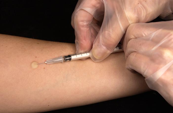
The Mantoux tuberculin skin test (TST) is the standard method of determining whether a person is infected with Mycobacterium tuberculosis. Reliable administration and reading of the TST requires standardization of procedures, training, supervision, and practice. The TST is performed by injecting 0.1 ml of tuberculin purified protein derivative (PPD) into the inner surface of the forearm. The injection should be made with a tuberculin syringe, with the needle bevel facing upward. The TST is an intradermal injection. When placed correctly, the injection should produce a pale elevation of the skin (a wheal) 6 to 10 mm in diameter.
The skin test reaction should be read between 48 and 72 hours after administration. A patient who does not return within 72 hours will need to be rescheduled for another skin test.
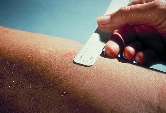
The reaction should be measured in millimeters of the induration (palpable, raised, hardened area or swelling). The reader should not measure erythema (redness). The diameter of the indurated area should be measured across the forearm (perpendicular to the long axis).
Classification of Tuberculin Reaction [1]


| Tuberculin Rection | Considered a Positive Result in: |
|---|---|
| ≥ 5 mm |
|
| ≥ 10 mm |
|
| ≥ 15 mm |
|
BCG Vaccine and Tuberculin Skin Test
There is disagreement on the use of the Mantoux test on people who have been immunized with BCG. The US recommendation is that in administering and interpreting the Mantoux test, previous BCG vaccination should be ignored; the UK recommendation is that interferon-γ tests should be used to help interpret positive tuberculin tests, also, the UK do not recommend serial tuberculin skin testing in people who have had BCG (a key part of the US strategy). In their guidelines on the use of QuantiFERON Gold the US Centers for Disease Control and Prevention state that whereas Quantiferon Gold is not affected by BCG inoculation tuberculin tests can be affected.[2] In general the US approach is likely to result in more false positives and more unnecessary treatment with potentially toxic drugs; the UK approach is as sensitive in theory and should also be more specific, because of the use of interferon-γ tests.
Under the US recommendations, latent TB infection (LTBI) diagnosis and treatment for LTBI is considered for any BCG-vaccinated person whose skin test is 10 mm or greater, if any of these circumstances are present:
- Was in contact with another person with infectious TB
- Was born or has resided in a high TB prevalence country
- Is continually exposed to populations where TB prevalence is high.
Microbiological Studies
A definitive diagnosis of tuberculosis can only be made by culturing Mycobacterium tuberculosis organisms from a specimen taken from the patient (most often sputum, but may also include pus, CSF, biopsied tissue, etc). A diagnosis made other than by culture may only be classified as probable or presumed.

Sputum smears and cultures should be done for acid-fast bacilli if the patient is producing sputum. The preferred method for this is fluorescence microscopy (auramine-rhodamine staining), which is more sensitive than conventional Ziehl-Neelsen staining.[3]
If no sputum is being produced, specimens can be obtained by inducing sputum, gastric washings, a laryngeal swab, bronchoscopy with bronchoalveolar lavage, or fine needle aspiration of a collection. A comparative study found that inducing three sputum samples is more sensitive than three gastric washings.[4]
Other mycobacteria are also acid-fast. If the smear is positive, PCR or gene probe tests can distinguish M. tuberculosis from other mycobacteria. Even if sputum smear is negative, tuberculosis must be considered and is only excluded after negative cultures.
Many types of cultures are available [5]. Traditionally, cultures have used the Löwenstein-Jensen (LJ), Kirchner, or Middlebrook media (7H9, 7H10, and 7H11). A culture of the AFB can distinguish the various forms of mycobacteria, although results from this may take four to eight weeks for a conclusive answer. New automated systems that are faster include the MB/BacT, BACTEC 9000, and the Mycobacterial Growth Indicator Tube (MGIT). The MODS culture may be a faster and more accurate method [6].
- Diagnostic Microbiology (sputum): The presence of acid-fast-bacilli (AFB) on a sputum smear or other specimen often indicates TB disease (at least 10,000c is needed on the smear to get a postive acid fast bacilli (AFB) stain). Acid-fast microscopy is easy and quick, but it does not confirm a diagnosis of TB because some acid-fast-bacilli are not M. tuberculosis. Therefore, a culture is done on all initial samples to confirm the diagnosis. However, a positive culture is not always necessary to begin or continue treatment for TB because cultures can take up to 3 weeks to yield definite results. A positive culture for M. tuberculosis confirms the diagnosis of TB disease. Culture examinations should be completed on all specimens, regardless of AFB smear results. Laboratories should report positive results on smears and cultures within 24 hours by telephone or fax to the primary health care provider and to the state or local TB control program, as required by law. A mycobacterium tuberculosis direct test (MTD) of nucleic acid amplification can also be performed to diagnose TB. An MTD test is similar to a polymerase chain reaction (pcr) and is very specific. The test is more sensitive than a smear but it is less senstive than a culture, and has the benefit of same day results.
- Diagnostic Microbiology (pleural fluid): A sample of pleural exudate can be analyzed by cytopathology or at a cell count lab. Samples are usually lymphocyte predominant, and cytopathology is more accurate than cell count labs at detecting lymphs. If there is more fluid present, then an AFB lab is more appropriate. A pleural exudate lab test may find sterile pyuria (especially in HIV positive patients), but overall this finding is fairly uncommon.
- Note: Most extra-pulmonary TB is pauci-bacillary, so the yield of tests is very low. This means that negative cultures do not mean no disease (e.g. negative cerebrospinal fluid AFB or even MTD is not that sensitive).
Heaf Test
The Heaf test is a diagnostic skin test performed in order to determine whether or not a child has been exposed to tuberculosis. Patients who exhibit a negative reaction to the test may be offered BCG vaccination. The test is named after F. R. G. Heaf.
Until 2005, the test was used in the United Kingdom to determine if the BCG vaccine was needed; the Mantoux test is now used instead. The Heaf test was preferred in the UK, because it was felt that the Heaf test was easier to interpret, with less inter-observer variability, and that less training was required to administer and to read the test. The test was withdrawn because manufacturers could not be found for tuberculin or for Heaf guns.
The Heaf test may be informally referred to as the six pricks, as it gives six individual injections.
Procedure
A Heaf gun is used to inject multiple samples of testing serum under the skin at once. A Heaf gun with disposable single-use heads is recommended.
The gun injects purified protein derivative equivalent to 100,000 units per mL to the skin over the flexor surface of the left forearm in a circular pattern of six. The test is read between 2 and 7 days later. The injection must not be into sites containing superficial veins.
The reading of the Heaf test is defined by a scale:
- Negative - No induration, maybe 6 minute puncture scars
- Grade 1 - 4-6 papules (also considered negative)
- Grade 2 - Confluent papules form indurated ring (positive)
- Grade 3 - Central filling to form disc (positive)
- Grade 4 - Disc >10 mm with or without blistering (strongly positive)
Grades 1 and 2 may be the result of previous BCG or avian tuberculosis.
Children who have a grade 3 or 4 reaction require X-ray and follow-up.
The equivalent Mantoux test positive levels done with 10 TU (0.1 mL 100 TU/mL, 1:1000) are:
- 0-4 mm induration (Heaf 0-1)
- 5-14 mm induration (Heaf 2)
- >15 mm induration (Heaf 3-4)
The Mantoux test is preferred in the United States for the diagnosis of tuberculosis; multiple puncture tests, such as the Heaf test and Tine test, are not recommended.
Drug Resistance
For all patients, the initial M. tuberculosis isolate should be tested for drug resistance. It is crucial to identify drug resistance as early as possible to ensure effective treatment. Drug susceptibility patterns should be repeated for patients who do not respond adequately to treatment or who have positive culture results despite 3 months of therapy. Susceptibility results from laboratories should be promptly reported to the primary health care provider and the state or local TB control program.
References
- ↑ "CDC Tuberculin Skin Testing".
- ↑ CDC - Guidelines for Using the QuantiFERON®-TB Gold Test for Detecting Mycobacterium tuberculosis Infection, United States
- ↑ Steingart K, Henry M, Ng V; et al. (2006). "Fluorescence versus conventional sputum smear microscopy for tuberculosis: a systematic review". Lancet Infect Dis. 6 (9): 570&ndash, 81. doi:10.1016/S1473-3099(06)70578-3.
- ↑ Brown M, Varia H, Bassett P, Davidson RN, Wall R, Pasvol G (2007). "Prospective study of sputum induction, gastric washing, and bronchoalveolar lavage for the diagnosis of pulmonary tuberculosis in patients who are unable to expectorate". Clin Infect Dis. 44 (11): 1415–20. doi:10.1086/516782. PMID 17479935.
- ↑ Drobniewski F, Caws M, Gibson A, Young D (2003). "Modern laboratory diagnosis of tuberculosis". Lancet Infect Dis. 3 (3): 141–7. PMID 12614730.
- ↑ Moore D, Evans C, Gilman R, Caviedes L, Coronel J, Vivar A, Sanchez E, Piñedo Y, Saravia J, Salazar C, Oberhelman R, Hollm-Delgado M, LaChira D, Escombe A, Friedland J (2006). "Microscopic-observation drug-susceptibility assay for the diagnosis of TB". N Engl J Med. 355 (15): 1539–50. PMID 17035648.