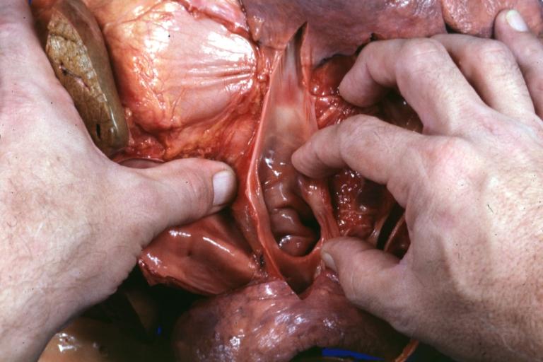
Total anomalous pulmonary venous connection
For patient information click here
| Total anomalous pulmonary venous connection | |
 | |
|---|---|
| Common pulmonary vein | |
| ICD-10 | Q26.2 |
| ICD-9 | 747.41 |
|
Total anomalous pulmonary venous connection Microchapters |
|
Differentiating Total anomalous pulmonary venous connection from other Diseases |
|---|
|
Diagnosis |
|
Treatment |
|
Case Studies |
|
Total anomalous pulmonary venous connection On the Web |
|
American Roentgen Ray Society Images of Total anomalous pulmonary venous connection |
|
Risk calculators and risk factors for Total anomalous pulmonary venous connection |
Editor-In-Chief: C. Michael Gibson, M.S., M.D. [1]
Associate Editors-In-Chief: Cafer Zorkun, M.D., Ph.D. [2]; Keri Shafer, M.D. [3]
Overview
Total anomalous pulmonary venous connection (TAPVC), also known as total anomalous pulmonary venous drainage(TAPVD) and total anamalous pulmonary venous return(TAPVR), is a rare cyanotic congenital heart defect (CHD) in which all four pulmonary veins are malpositioned and make anomalous connections to the systemic venous circulation. (Normally, pulmonary venous return carries oxygenated blood to the left atrium and to the rest of the body). A patent foramen ovale or an atrial septal defect must be present in order to allow systemic blood flow.
Variations
There are four variants:
- Supracardiac (50%): blood drains to one of the innominate veins (brachiocephalic veins) or the superior vena cava
- Cardiac (20%): blood drains into coronary sinus or directly into right atrium
- Infradiaphragmatic (20%): blood drains into portal or hepatic veins
- Mixed (10%)
TAPVC can occur with obstruction, which occurs when the anomalous vein enters a vessel at an acute angle and can cause pulmonary venous hypertension and cyanosis because blood cannot easily enter the new vein as easily.
Diagnosis
Symptoms
- cyanosis, tachypnea, dyspnea since the overloaded pulmonary circuit can cause pulmonary edema
Physical Examination
- right ventricular heave
- fixed split S2
- S3 gallop
- systolic ejection murmur at left upper sternal border
- cardiomegaly
Electrocardiographic Findings
Cardiac Catheterization
<googlevideo>4462444442806796487&hl=en</googlevideo>
<googlevideo>-3314937685295122195&hl=en</googlevideo>
<googlevideo>2240824856713063576&pr=goog-sl</googlevideo>
<googlevideo>4846170916216525758&hl=en</googlevideo>
<googlevideo>2692134944789571414&hl=en</googlevideo>
<googlevideo>961264589763540810&hl=en</googlevideo>
<googlevideo>613495638748988173&hl=en</googlevideo>
Treatment
In TAPVC without obstruction, surgical redirection can be performed within the first month of life. With obstruction, surgery should be undertaken emergently. PGE1 should not be given because a patent ductus arteriosus adds even more volume into the already overloaded pulmonary flow.
References
External links
- Total Anomalous Pulmonary Venous Return information from Seattle Children's Hospital Heart Center
Additional Reading
- Moss and Adams' Heart Disease in Infants, Children, and Adolescents Hugh D. Allen, Arthur J. Moss, David J. Driscoll, Forrest H. Adams, Timothy F. Feltes, Robert E. Shaddy, 2007 ISBN 0781786843