Sandbox/NCT
Characterize the symptoms:
| |||||||||||||||||||||||||||||||||||||||||||||||
Identify possible triggers: | |||||||||||||||||||||||||||||||||||||||||||||||
Differential Diagnosis | |||||||||||||||||||||||||||||||||||||||||||||||
Examine the patient: General appearance Vitals
Skin
Neck
Cardiovascular examination
| |||||||||||||||||||||||||||||||||||||||||||||||
❑ Assess hemodynamic stability
❑ Order and monitor the ECG | |||||||||||||||||||||||||||||||||||||||||||||||
| ❑ Unstable patient | ❑ Stable patient | ||||||||||||||||||||||||||||||||||||||||||||||
❑ If the rythm isn't sinus tachycardia: Urgent cardioversion | ❑ If the rythm is sinus tachycardia: Focus your treatment on the underlying condition. If it is due to cardiac ischemia or aortic stenosis, control heart rate by IV metoprolol at the rate of 5 mg/2 minutes till full control or till the maximum of 15 mg, then shift to oral regimen. Don't adminster beta blockers if the patient has significant bradycardia (<50 beats per minute) | Documented arrhythmia | Undocumented arrhythmia (ECG is normal) | ||||||||||||||||||||||||||||||||||||||||||||
❑ Confirm diagnosis of narrow QRS complex tachycardia (heart rate > 100 beats per minute associated with a QRS complex duration < 120 milliseconds) ❑ Identify and treat SVT | History suggestive of extra premature beats ❑ Sensation of a pause followed by a strong heart beat OR | History suggestive of paroxysmal arrhythmia ❑ Regular palpitations with sudden onset and termination | |||||||||||||||||||||||||||||||||||||||||||||
❑ Refer for an invasive electrophysiological study AND/OR ❑ Catheter ablation ❑ Educate about vagal maneuvers ❑ Consider beta blocker | |||||||||||||||||||||||||||||||||||||||||||||||
Differential Diagnosis
| Type of Arrhythmia | EKG (lead II)† | Clues |
| Supraventricular tachycardia | 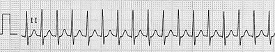 |
Any tachyarrhythmia that is initiated and maintained in atrial tissue or atrioventricular junctional tissue.[1] |
| Sinus tachycardia |  |
Rhythm with heart rate > 100 bpm, originating in SA node due to its increased automaticity. |
| Sinus node re-entry tachycardia | Rare paroxysmal tachycardia arising due to re-entry circuits with in SA node.[2] | |
| Atrial fibrillation |  |
Supraventricular tachycardia with irregularly irregular rhythm and absent P waves on EKG. |
| Atrial flutter |  |
Cardiac rhythm characterized by an atrial rate ranging from 240 to 400 beats per minute and regular continuous wave-form.[3] |
| AVNRT |  |
Most common form of PSVT with a heart rate of 140-250 bpm, re-entrant circuit involves two separate anatomical pathways (slow and fast) loacted in perinodal tissue. |
| AVRT |  |
Re-entrant tachycardia occurring due to an accessory pathway in addition to AV node, accessory pathway is essential for the initiation and the maintenance of tachycardia. |
| Focal atrial tachycardia |  |
Focal atria tachycardia refers to a rhythm originating from a single site either in the left or right atrium with an atrial rate of 100-250 bpm. |
| Nonparoxysmal junctional tachycardia |  |
Benign tachycardia occurring due to increased automaticity arising from a high junctional focus. |
| Multifocal atrial tachycardia |  |
Irregular tachycardia characterized by 3 different P wave morphologies on EKG. |
† EKG strips is a courtesy from ECGpedia.
References
- ↑ "ACC/AHA/ESC Guidelines for the Management of Patients With Supraventricular Arrhythmias—Executive Summary". Retrieved 15 August 2013.
- ↑ Cossú, SF.; Steinberg, JS. "Supraventricular tachyarrhythmias involving the sinus node: clinical and electrophysiologic characteristics". Prog Cardiovasc Dis. 41 (1): 51–63. PMID 9717859.
- ↑ Dhar S, Lidhoo P, Koul D, Dhar S, Bakhshi M, Deger FT (2009). "Current concepts and management strategies in atrial flutter". South. Med. J. 102 (9): 917–22. doi:10.1097/SMJ.0b013e3181b0f4b8. PMID 19668035. Unknown parameter
|month=ignored (help)