Pneumonia natural history, complications, and prognosis
|
Pneumonia Microchapters |
|
Diagnosis |
|---|
|
Treatment |
|
Case Studies |
|
Pneumonia natural history, complications, and prognosis On the Web |
|
American Roentgen Ray Society Images of Pneumonia natural history, complications, and prognosis |
|
FDA on Pneumonia natural history, complications, and prognosis |
|
CDC onPneumonia natural history, complications, and prognosis |
|
Pneumonia natural history, complications, and prognosis in the news |
|
Blogs on Pneumonia natural history, complications, and prognosis |
|
Risk calculators and risk factors for Pneumonia natural history, complications, and prognosis |
Editor-In-Chief: C. Michael Gibson, M.S., M.D. [1] ;Associate Editor(s)-in-Chief: Priyamvada Singh, M.D. [2]
Complications
Despite appropriate antibiotic therapy, severe complications can result from CAP, including:
Sepsis
- Sepsis can occur when microorganisms enter the blood stream and the immune system responds.
- Sepsis most often occurs with bacterial pneumonia
- Streptococcus pneumoniae is the most common cause.
- Individuals with sepsis require hospitalization in an intensive care unit. They often require medications and intravenous fluids to keep their blood pressure from going too low. Sepsis can cause liver, kidney, and heart damage among other things.
Respiratory Failure
- If enough of the lung is involved, it may not be possible for a person to breathe enough to live without support.
- Non-invasive machines such as a bilevel positive airway pressure machine may be used.
- Otherwise, placement of a breathing tube into the mouth may be necessary. A ventilator may also be used to help the person breathe.
Pleural Effusion and Empyema
- Occasionally, microorganisms from the lung will cause fluid to form in the space surrounding the lung, called the pleural cavity.
- If the microorganisms themselves are present, the fluid collection is often called an empyema.
- If pleural fluid is present in a person with CAP, the fluid should be collected with a needle (thoracentesis) and examined.
- Depending on the result of the examination, complete drainage of the fluid may be necessary, often with a chest tube. If the fluid is not drained, bacteria can continue to cause illness because antibiotics do not penetrate well into the pleural cavity.
Abscess
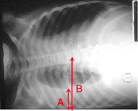
- Rarely, microorganisms in the lung will form a pocket of fluid and bacteria called an abscess.
- Abscesses can be seen on an x-ray as a cavity within the lung. Abscesses typically occur in aspiration pneumonia and most often contain a mixture of anaerobic bacteria.
- Usually antibiotics are able to fully treat abscesses, but sometimes they must be drained by a surgeon or radiologist.
Sometimes pneumonia can lead to additional complications. Complications are more frequently associated with bacterial pneumonia than with viral pneumonia. The most important complications include:
Respiratory and Circulatory Failure
Because pneumonia affects the lungs, often people with pneumonia have difficulty breathing, and it may not be possible for them to breathe well enough to stay alive without support. Non-invasive breathing assistance may be helpful, such as with a bilevel positive airway pressure machine. In other cases, placement of an endotracheal tube (breathing tube) may be necessary, and a ventilator may be used to help the person breathe.
Pneumonia can also cause respiratory failure by triggering acute respiratory distress syndrome (ARDS), which results from a combination of infection and inflammatory response. The lungs quickly fill with fluid and become very stiff. This stiffness, combined with severe difficulties extracting oxygen due to the alveolar fluid, creates a need for mechanical ventilation.

Sepsis and septic shock are potential complications of pneumonia. Sepsis occurs when microorganisms enter the bloodstream and the immune systemresponds by secreting cytokines. Sepsis most often occurs with bacterial pneumonia; Streptococcus pneumoniae is the most common cause. Individuals with sepsis or septic shock need hospitalization in an intensive care unit. They often require intravenous fluids and medications to help keep their blood pressure from dropping too low. Sepsis can cause liver, kidney, and heart damage, among other problems, and it often causes death.
Pleural Effusion, Empyema, and Abscess
Occasionally, microorganisms infecting the lung will cause fluid (a pleural effusion) to build up in the space that surrounds the lung (the pleural cavity). If the microorganisms themselves are present in the pleural cavity, the fluid collection is called an empyema. When pleural fluid is present in a person with pneumonia, the fluid can often be collected with a needle (thoracentesis) and examined. Depending on the results of this examination, complete drainage of the fluid may be necessary, often requiring a chest tube. In severe cases of empyema, surgery may be needed. If the fluid is not drained, the infection may persist, because antibiotics do not penetrate well into the pleural cavity.
Rarely, bacteria in the lung will form a pocket of infected fluid called an abscess. Lung abscesses can usually be seen with a chest x-ray or chest CT scan. Abscesses typically occur in aspiration pneumonia and often contain several types of bacteria. Antibiotics are usually adequate to treat a lung abscess, but sometimes the abscess must be drained by a surgeon or radiologist.
Prognosis
- Individuals who are treated for CAP outside of the hospital have a mortality rate less than 1%.
- Fever typically responds in the first two days of therapy and other symptoms resolve in the first week.
- The x-ray, however, may remain abnormal for at least a month, even when CAP has been successfully treated.
- Among individuals who require hospitalization, the mortality rate averages 12% overall, but it is as much as 40% in people who have bloodstream infections or require intensive care.[3]
- When CAP does not respond as expected, there are several possible causes.
- A complication of CAP may have occurred or a previously unknown health problem may be playing a role.
- Additional causes include inappropriate antibiotics for the causative organism (ie DRSP), a previously unsuspected microorganism (such as tuberculosis), or a condition which mimics CAP (such as Wegener's granulomatosis).
- Additional testing may be performed and may include additional radiologic imaging (such as a computed tomography scan) or a procedure such as a bronchoscopy or lung biopsy.
Prognosis and Mortality
With treatment, most types of bacterial pneumonia can be cured within one to two weeks. Viral pneumonia may last longer, and mycoplasmal pneumonia may take four to six weeks to resolve completely. The eventual outcome of an episode of pneumonia depends on how ill the person is when he or she is first diagnosed.
In the United States, about one of every twenty people with pneumococcal pneumonia will die.[1] In cases where the pneumonia progresses to blood poisoning (bacteremia), one of every five will die. The death rate (or mortality) also depends on the underlying cause of the pneumonia. Pneumonia caused by Mycoplasma, for instance, is associated with little mortality. However, about half of the people who develop methicillin-resistantStaphylococcus aureus (MRSA) pneumonia while on a ventilator will die.[2] In regions of the world without advanced health care systems, pneumonia is even deadlier. Limited access to clinics and hospitals, limited access to x-rays, limited antibiotic choices, and inability to treat underlying conditions inevitably leads to higher rates of death from pneumonia.
Clinical prediction rules
Clinical prediction rules have been developed to more objectively prognosticate outcomes in pneumonia. These rules can be helpful in deciding whether or not to hospitalize the person.
- Pneumonia severity index[3] -online calculator
- CURB-65 score, which takes into account the severity of symptoms, any underlying diseases, and age.[4] -online calculator
References
- ↑ http://www.kidshealth.org/parent/infections/bacterial_viral/pneumonia.html
- ↑ Combes A, Luyt CE, Fagon JY, Wollf M, Trouillet JL, Gibert C, Chastre J; PNEUMA Trial Group. Impact of methicillin resistance on outcome of Staphylococcus aureus ventilator-associated pneumonia. Am J Respir Crit Care Med. 2004 Oct 1;170(7):786-92. PMID 15242840
- ↑ Fine MJ, Auble TE, Yealy DM, Hanusa BH, Weissfeld LA, Singer DE, Coley CM, Marrie TJ, Kapoor WN. A prediction rule to identify low-risk patients with community-acquired pneumonia. N Engl J Med. 1997 Jan 23;336(4):243–250. PMID 8995086
- ↑ Lim WS, van der Eerden MM, Laing R; et al. (2003). "Defining community acquired pneumonia severity on presentation to hospital: an international derivation and validation study". Thorax. 58 (5): 377–82. PMID 12728155.