Nonossifying fibroma: Difference between revisions
Hudakarman (talk | contribs) (→X-ray) |
Hudakarman (talk | contribs) |
||
| (One intermediate revision by the same user not shown) | |||
| Line 47: | Line 47: | ||
***[[Congenital syndromes|Congenital syndrome]] of multiple non-ossifying fibromas and [[Café au lait spot|cafe au lait]] [[pigmentation]], [[mental retardation]], [[heart]], [[eyes]] and [[gonads]] ([[hypogonadism]]) involved | ***[[Congenital syndromes|Congenital syndrome]] of multiple non-ossifying fibromas and [[Café au lait spot|cafe au lait]] [[pigmentation]], [[mental retardation]], [[heart]], [[eyes]] and [[gonads]] ([[hypogonadism]]) involved | ||
**[[Neurofibromatosis]] | **[[Neurofibromatosis]] | ||
**Familial multifocal NOF | **[[Familial]] multifocal NOF | ||
**[[Aneurysmal bone cyst]] | **[[Aneurysmal bone cyst]] | ||
| Line 128: | Line 128: | ||
==Natural History, Complications, and Prognosis== | ==Natural History, Complications, and Prognosis== | ||
*Common complications of [[Aneurysmal disease|aneurysmal]] bone cyst include:<ref>{{cite journal |vauthors=Wodajo FM |title=Top five lesions that do not need referral to orthopedic oncology |journal=Orthop. Clin. North Am. |volume=46 |issue=2 |pages=303–14 |date=April 2015 |pmid=25771324 |doi=10.1016/j.ocl.2014.11.012 |url=}}</ref> | *Common [[complications]] of [[Aneurysmal disease|aneurysmal]] [[bone cyst]] include:<ref>{{cite journal |vauthors=Wodajo FM |title=Top five lesions that do not need referral to orthopedic oncology |journal=Orthop. Clin. North Am. |volume=46 |issue=2 |pages=303–14 |date=April 2015 |pmid=25771324 |doi=10.1016/j.ocl.2014.11.012 |url=}}</ref> | ||
**[[Pathological]] [[Bone fracture|fracture]] | **[[Pathological]] [[Bone fracture|fracture]] | ||
**Premature [[Epiphyseal plate|epiphyseal]] closure | **Premature [[Epiphyseal plate|epiphyseal]] closure | ||
| Line 149: | Line 149: | ||
*[[X-rays|X-ray]] is the diagnostic study of choice for the diagnosis of non ossifying fibroma. | *[[X-rays|X-ray]] is the diagnostic study of choice for the diagnosis of non ossifying fibroma. | ||
*[[X-rays|X-ray]] findings include:<ref>{{cite journal |vauthors=Ritschl P, Karnel F, Hajek P |title=Fibrous metaphyseal defects--determination of their origin and natural history using a radiomorphological study |journal=Skeletal Radiol. |volume=17 |issue=1 |pages=8–15 |date=1988 |pmid=3358140 |doi= |url=}}</ref><ref>{{cite journal |vauthors=Drennan DB, Maylahn DJ, Fahey JJ |title=Fractures through large non-ossifying fibromas |journal=Clin. Orthop. Relat. Res. |volume= |issue=103 |pages=82–8 |date=1974 |pmid=4413505 |doi= |url=}}</ref><ref>{{cite journal |vauthors=Ritschl P, Karnel F, Hajek P |title=Fibrous metaphyseal defects--determination of their origin and natural history using a radiomorphological study |journal=Skeletal Radiol. |volume=17 |issue=1 |pages=8–15 |date=1988 |pmid=3358140 |doi= |url=}}</ref> | *[[X-rays|X-ray]] findings include:<ref>{{cite journal |vauthors=Ritschl P, Karnel F, Hajek P |title=Fibrous metaphyseal defects--determination of their origin and natural history using a radiomorphological study |journal=Skeletal Radiol. |volume=17 |issue=1 |pages=8–15 |date=1988 |pmid=3358140 |doi= |url=}}</ref><ref>{{cite journal |vauthors=Drennan DB, Maylahn DJ, Fahey JJ |title=Fractures through large non-ossifying fibromas |journal=Clin. Orthop. Relat. Res. |volume= |issue=103 |pages=82–8 |date=1974 |pmid=4413505 |doi= |url=}}</ref><ref>{{cite journal |vauthors=Ritschl P, Karnel F, Hajek P |title=Fibrous metaphyseal defects--determination of their origin and natural history using a radiomorphological study |journal=Skeletal Radiol. |volume=17 |issue=1 |pages=8–15 |date=1988 |pmid=3358140 |doi= |url=}}</ref> | ||
**[[Metaphyseal]] eccentric "bubbly" lytic lesion surrounded by sclerotic rim | **[[Metaphyseal]] eccentric "bubbly" [[lytic]] [[lesion]] surrounded by sclerotic rim | ||
**[[Cortex]] may be expanded and thin | **[[Cortex]] may be expanded and thin | ||
**As bone grows, it migrates to [[diaphysis]] and the lesions enlarge. | **As bone grows, it migrates to [[diaphysis]] and the lesions enlarge. | ||
| Line 155: | Line 155: | ||
===History and Symptoms=== | ===History and Symptoms=== | ||
*The majority of patients with non ossifying fibroma are asymptomatic.<ref>{{cite book | last = Peabody | first = Terrance | title = Orthopaedic oncology : primary and metastatic tumors of the skeletal system | publisher = Springer | location = Cham | year = 2014 | isbn = 9783319073224 }}</ref> | *The majority of patients with non ossifying fibroma are [[asymptomatic]].<ref>{{cite book | last = Peabody | first = Terrance | title = Orthopaedic oncology : primary and metastatic tumors of the skeletal system | publisher = Springer | location = Cham | year = 2014 | isbn = 9783319073224 }}</ref> | ||
*Some patients with non ossifying fibroma have a positive history of: | *Some [[Patient|patients]] with non ossifying fibroma have a positive [[History and Physical examination|history]] of: | ||
**[[Pain]] | **[[Pain]] | ||
**[[Swelling]] | **[[Swelling]] | ||
| Line 164: | Line 164: | ||
*Patients with non ossifying fibroma usually appear well. | *Patients with non ossifying fibroma usually appear well. | ||
*Common [[physical examination]] findings of non ossifying fibroma include:<ref>{{cite journal |vauthors=Mallet JF, Rigault P, Padovani JP, Touzet P, Nezelof C |title=[Non-ossifying fibroma in children: a surgical condition?] |language=French |journal=Chir Pediatr |volume=21 |issue=3 |pages=179–89 |date=1980 |pmid=7408072 |doi= |url=}}</ref><ref>{{cite journal |vauthors=Fauré C, Laurent JM, Schmit P, Sirinelli D |title=Multiple and large non-ossifying fibromas in children with neurofibromatosis |journal=Ann Radiol (Paris) |volume=29 |issue=3-4 |pages=369–73 |date=1986 |pmid=3092720 |doi= |url=}}</ref><ref>{{cite journal |vauthors=Hetts SW, Hilchey SD, Wilson R, Franc B |title=Case 110: Nonossifying fibroma |journal=Radiology |volume=243 |issue=1 |pages=288–92 |date=April 2007 |pmid=17392261 |doi=10.1148/radiol.2431040427 |url=}}</ref> | *Common [[physical examination]] findings of non ossifying fibroma include:<ref>{{cite journal |vauthors=Mallet JF, Rigault P, Padovani JP, Touzet P, Nezelof C |title=[Non-ossifying fibroma in children: a surgical condition?] |language=French |journal=Chir Pediatr |volume=21 |issue=3 |pages=179–89 |date=1980 |pmid=7408072 |doi= |url=}}</ref><ref>{{cite journal |vauthors=Fauré C, Laurent JM, Schmit P, Sirinelli D |title=Multiple and large non-ossifying fibromas in children with neurofibromatosis |journal=Ann Radiol (Paris) |volume=29 |issue=3-4 |pages=369–73 |date=1986 |pmid=3092720 |doi= |url=}}</ref><ref>{{cite journal |vauthors=Hetts SW, Hilchey SD, Wilson R, Franc B |title=Case 110: Nonossifying fibroma |journal=Radiology |volume=243 |issue=1 |pages=288–92 |date=April 2007 |pmid=17392261 |doi=10.1148/radiol.2431040427 |url=}}</ref> | ||
**Asymptomatic | **[[Asymptomatic]] | ||
**[[Pain]] if associated with [[pathological]] [[fracture]] | **[[Pain]] if associated with [[pathological]] [[fracture]] | ||
*Findings associated with [[Jaffe-Campanacci syndrome]] include:<ref>{{cite journal |vauthors=Cherix S, Bildé Y, Becce F, Letovanec I, Rüdiger HA |title=Multiple non-ossifying fibromas as a cause of pathological femoral fracture in Jaffe-Campanacci syndrome |journal=BMC Musculoskelet Disord |volume=15 |issue= |pages=218 |date=June 2014 |pmid=24965055 |pmc=4088300 |doi=10.1186/1471-2474-15-218 |url=}}</ref> | *Findings associated with [[Jaffe-Campanacci syndrome]] include:<ref>{{cite journal |vauthors=Cherix S, Bildé Y, Becce F, Letovanec I, Rüdiger HA |title=Multiple non-ossifying fibromas as a cause of pathological femoral fracture in Jaffe-Campanacci syndrome |journal=BMC Musculoskelet Disord |volume=15 |issue= |pages=218 |date=June 2014 |pmid=24965055 |pmc=4088300 |doi=10.1186/1471-2474-15-218 |url=}}</ref> | ||
Latest revision as of 20:55, 9 October 2019
Editor-In-Chief: C. Michael Gibson, M.S., M.D. [1]; Associate Editor(s)-in-Chief: Rohan A. Bhimani, M.B.B.S., D.N.B., M.Ch.[2] Huda A. Karman, M.D.
Synonyms and keywords: Fibroxanthoma; Fibrous cortical defect; NOF
Overview
Non ossifying fibromas are present in about 30 % of children. The incidence of Non ossifying fibroma is approximately 1000-2000 per 100,000 individuals worldwide. The age distribution of Non ossifying fibroma is between 5-15 years. Men are more commonly affected than women, with a 1.9:1 ratio. In 1929, Phemister first described the term non ossifying fibroma. The exact pathogenesis of non ossifying fibroma is not fully understood. Non ossifying fibroma (NOF) typically occur in the metaphysis of the long bones. The bones often involved are femur, tibia, and fibula. The majority of patients with non ossifying fibroma are asymptomatic. Some patients with non ossifying fibroma have a complaints of pain, swelling and pathological fracture. X-ray is the diagnostic study of choice for the diagnosis of non ossifying fibroma demonstrating soap bubble like lytic lesion. Observation is the mainstay of treatment for non ossifying fibroma. Surgery in form of curettage and bone grafting is reserved for cases with high risk of pathological fracture.
Historical Perspective
- In 1929, Phemister first described the term non ossifying fibroma.[1][2]
- In 1941, Sontag and Pyle reported a radiologic description of non ossifying fibroma.
- In 1942, Jaffe and Lichtenstein described clinical findings, anatomic aspects and the natural history.
Classification
Non ossifying fibroma can be classified based on imaging findings.
Enneking (MSTS) Staging System
- The Enneking surgical staging system (also known as the MSTS system) for benign musculoskeletal tumors based on radiographic characteristics of the tumor host margin.[3]
- It is widely accepted and routinely used classification.
| Stages | Description |
|---|---|
| 1 | Latent: Well demarcated borders |
| 2 | Active: Indistinct borders |
| 3 | Aggressive: Indistinct borders |
Pathophysiology
- The exact pathogenesis of non ossifying fibroma is not fully understood.
- Various theories have been proposed concerning the pathogenesis of non ossifying fibroma[4][5]
- An abnormal development extending from the growth plate.
- An abnormal osteoclastic resorption at the subperiosteal level during remodeling of the metaphysis.
- Non ossifying fibroma (NOF) typically occur in the metaphysis of the long bones.[6]
- The bones often involved are femur, tibia, and fibula.[7]
- Conditions associated:
- Jaffe-Campanacci syndrome
- Congenital syndrome of multiple non-ossifying fibromas and cafe au lait pigmentation, mental retardation, heart, eyes and gonads (hypogonadism) involved
- Neurofibromatosis
- Familial multifocal NOF
- Aneurysmal bone cyst
- Jaffe-Campanacci syndrome
Causes
Differentiating Non Ossifying Fibroma from Other Diseases
Non ossifying fibroma must be differentiated from following bone disorders:
| Disease | Bubbly lytic lesion on x-ray | Lakes of Blood on histology | Diagnosis | Treatment is curretage and bone grafting |
|---|---|---|---|---|
| Non ossifying fibroma | + | - | Radiology and biopsy | - |
| Unicameral bone cyst | + | - | Radiology and biopsy | - |
| Aneurysmal bone cyst | + | + | Radiology and biopsy | + |
| Giant cell tumor | - | - | Radiology and Biopsy | + |
| Chondroblastoma | - | - | Biopsy | + |
| Chondromyxoid Fibroma | - | - | Radiology and biopsy | + |
| Osteoblastoma | - | - | Radiology and biopsy | + |
| Telangiectatic osteosarcoma | - | + | Radiology and biopsy | - |
Epidemiology and Demographics
- The incidence of Non ossifying fibroma is approximately 1000 - 2000 per 100,000 individuals worldwide.[9]
- Adolescents and children are most affected by non ossifying fibroma.
- Non ossifying fibromas are present in about 30 % of children.[10]
- The age distribution of Non ossifying fibroma is between 5-15 years.
- Men are more commonly affected than women, with a 1.9:1 ratio.[11]
- There is no racial predilection to non ossifying fibroma.
Risk Factors
- There are no established risk factors for non ossifying fibroma.
Screening
- There is insufficient evidence to recommend routine screening for non ossifying fibroma.
Natural History, Complications, and Prognosis
- Common complications of aneurysmal bone cyst include:[12]
- Pathological fracture
- Premature epiphyseal closure
- Limb-length discrepancy
- Angular deformity
- Rare malignant transformation
- Prognosis is generally excellent for non ossifying fibroma.
- Factors that influence the outcome of the non ossifying fibroma include:
- Young age
- Open growth plates
- Metaphyseal location
Diagnosis
Diagnostic Study of Choice
 |
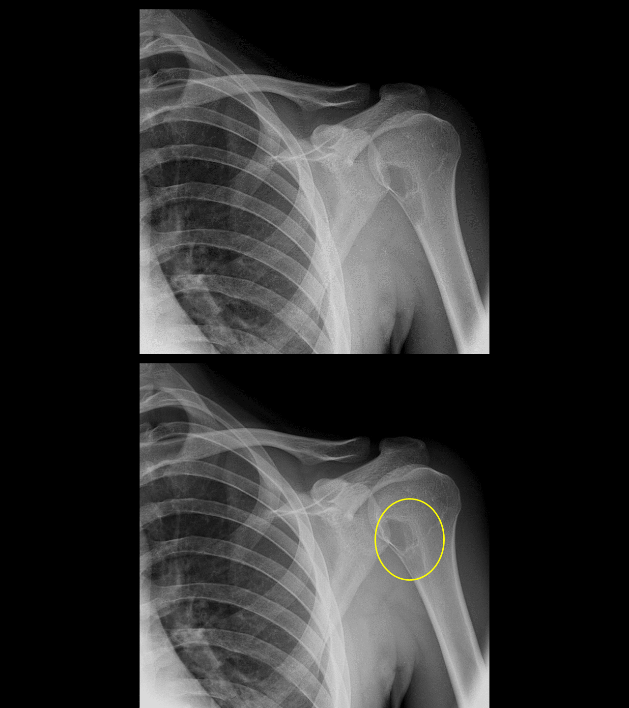
- X-ray is the diagnostic study of choice for the diagnosis of non ossifying fibroma.
- X-ray findings include:[13][14][15]
History and Symptoms
- The majority of patients with non ossifying fibroma are asymptomatic.[16]
- Some patients with non ossifying fibroma have a positive history of:
Physical Examination
- Patients with non ossifying fibroma usually appear well.
- Common physical examination findings of non ossifying fibroma include:[17][18][19]
- Asymptomatic
- Pain if associated with pathological fracture
- Findings associated with Jaffe-Campanacci syndrome include:[20]
- Café-au-lait spots
- Multiple nevi
- Hypogonadism
- Ocular abnormalities
Laboratory Findings
- There are no diagnostic laboratory findings associated with non ossifying fibroma.
Electrocardiogram
- There are no ECG findings associated with non ossifying fibroma.
X-ray
 |
Echocardiography or Ultrasound
- There are no echocardiography/ultrasound findings associated with non ossifying fibroma.
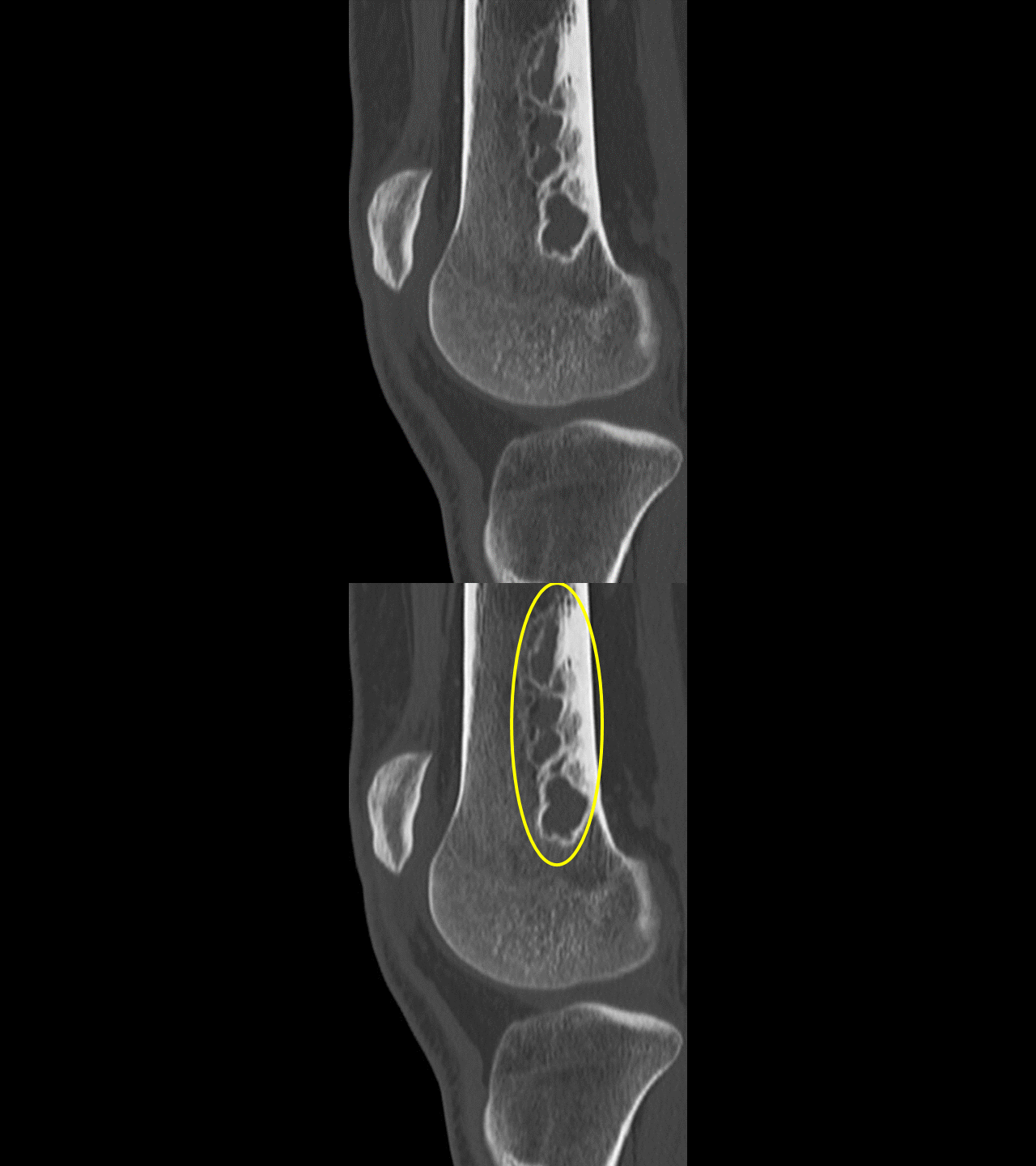
CT scan
- Computed tomography (CT) is useful in predicting the fracture risk.[24][25][26]
- CT scans may depict a central lucency.
- CT scan may confirm a minimally displaced fracture.
- It may also be helpful in preoperative planning in unusual locations such as the femoral neck.
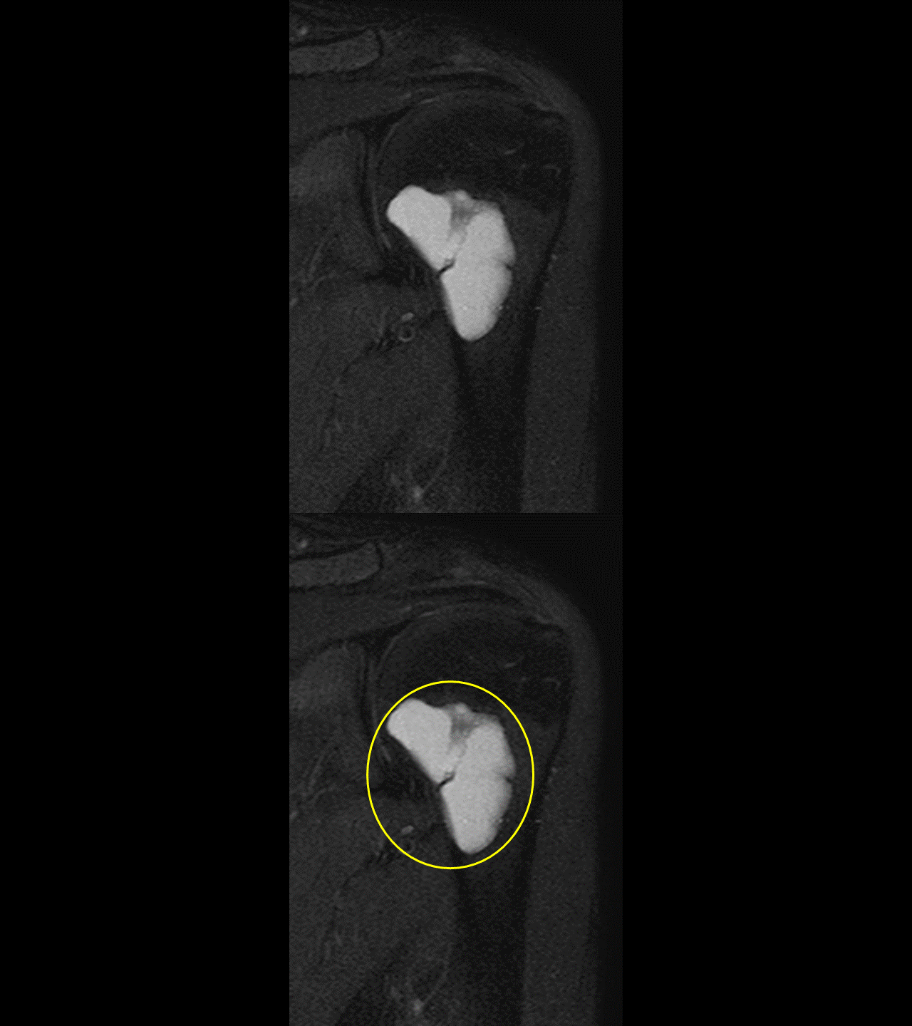
MRI
- Magnetic resonance imaging (MRI) are similar to those from CT scan.[27]
- MRI demonstrates a characteristic low signal on both T1 and T2 sequences without enhancement.
- High signal on T2 can be seen with an associated stress fracture.
Other Imaging Findings
- There are no other imaging findings associated with non ossifying fibroma.
 |
Other Diagnostic Studies
Biopsy
- Biopsy finding of non ossifying fibroma includes:[28]
- Bland fibroblastic component with a few histiocytes, myofibroblast cells, and giant cells.
- Marked proliferations of spindle cells arranged in a storiform pattern.
- Hemosiderin deposits also are found.
- Some leukocyte infiltration may be present.
Treatment
Medical Therapy
- Observation is the mainstay of treatment for non ossifying fibroma.[29]
Observation
Indications
- First line of treatment
- Most lesions resolve spontaneously and progressively re-ossify as child enters 2nd and 3rd decade of life
Technique
- Radiographs at 6, 12months, then annually until reossified
Casting
Indication
- Pathologic fracture especially in the pediatric population.
Surgery
Indication
- Unstable fractures
- High risk of pathologic fracture
Technique
- The surgical approach involves exposing the fracture site and developing a cortical window to curette the tumor.[30][31]
Primary Prevention
- There are no established measures for the primary prevention of non ossifying fibroma.
Secondary Prevention
- There are no established measures for the secondary prevention of non ossifying fibroma.
References
- ↑ H. L. Jaffe & L. Lichtenstein (1942). "Non-osteogenic fibroma of bone". The American journal of pathology. 18 (2): 205–221. PMID 19970624. Unknown parameter
|month=ignored (help) - ↑ Peabody, Terrance (2014). Orthopaedic oncology : primary and metastatic tumors of the skeletal system. Cham: Springer. ISBN 9783319073224.
- ↑ Jawad MU, Scully SP (2010). "In brief: classifications in brief: enneking classification: benign and malignant tumors of the musculoskeletal system". Clin Orthop Relat Res. 468 (7): 2000–2. doi:10.1007/s11999-010-1315-7. PMC 2882012. PMID 20333492.
- ↑ Herget GW, Mauer D, Krauß T, El Tayeh A, Uhl M, Südkamp NP, Hauschild O (April 2016). "Non-ossifying fibroma: natural history with an emphasis on a stage-related growth, fracture risk and the need for follow-up". BMC Musculoskelet Disord. 17: 147. doi:10.1186/s12891-016-1004-0. PMC 4820930. PMID 27044378.
- ↑ Goldin A, Muzykewicz DA, Dwek J, Mubarak SJ (October 2017). "The aetiology of the non-ossifying fibroma of the distal femur and its relationship to the surrounding soft tissues". J Child Orthop. 11 (5): 373–379. doi:10.1302/1863-2548.11.170068. PMC 5643931. PMID 29081852.
- ↑ Mankin HJ, Trahan CA, Fondren G, Mankin CJ (May 2009). "Non-ossifying fibroma, fibrous cortical defect and Jaffe-Campanacci syndrome: a biologic and clinical review". Chir Organi Mov. 93 (1): 1–7. doi:10.1007/s12306-009-0016-4. PMID 19711155.
- ↑ CUNNINGHAM JB, ACKERMAN LV (July 1956). "Metaphyseal fibrous defects". J Bone Joint Surg Am. 38-A (4): 797–808. PMID 13331975.
- ↑ Hatcher CH (December 1945). "The Pathogenesis of Localized Fibrous Lesions in the Metaphyses of Long Bones". Ann. Surg. 122 (6): 1016–30. PMC 1618342. PMID 17858695.
- ↑ Freyschmidt J, Ostertag H, Saure D (April 1981). "[Fibrous metaphyseal defect (fibrous cortical defect, non-ossifying fibroma). Paper II: differential diagnosis (author's transl)]". Rofo (in German). 134 (4): 392–400. doi:10.1055/s-2008-1056377. PMID 6453054.
- ↑ Nielsen GP, Kyriakos M. Fibrohistiocytic tumours. In: Fletcher CDM, Bridge J, Hogendorn PCW, Mertens F, editors. WHO Classifications of tumours of bone and soft Tissue. Lyon: IARC Press; 2013. pp. 301–4
- ↑ Ritschl P, Lintner F, Pechmann U, Brand G (1990). "Fibrous metaphyseal defect". Int Orthop. 14 (2): 205–11. PMID 2115506.
- ↑ Wodajo FM (April 2015). "Top five lesions that do not need referral to orthopedic oncology". Orthop. Clin. North Am. 46 (2): 303–14. doi:10.1016/j.ocl.2014.11.012. PMID 25771324.
- ↑ Ritschl P, Karnel F, Hajek P (1988). "Fibrous metaphyseal defects--determination of their origin and natural history using a radiomorphological study". Skeletal Radiol. 17 (1): 8–15. PMID 3358140.
- ↑ Drennan DB, Maylahn DJ, Fahey JJ (1974). "Fractures through large non-ossifying fibromas". Clin. Orthop. Relat. Res. (103): 82–8. PMID 4413505.
- ↑ Ritschl P, Karnel F, Hajek P (1988). "Fibrous metaphyseal defects--determination of their origin and natural history using a radiomorphological study". Skeletal Radiol. 17 (1): 8–15. PMID 3358140.
- ↑ Peabody, Terrance (2014). Orthopaedic oncology : primary and metastatic tumors of the skeletal system. Cham: Springer. ISBN 9783319073224.
- ↑ Mallet JF, Rigault P, Padovani JP, Touzet P, Nezelof C (1980). "[Non-ossifying fibroma in children: a surgical condition?]". Chir Pediatr (in French). 21 (3): 179–89. PMID 7408072.
- ↑ Fauré C, Laurent JM, Schmit P, Sirinelli D (1986). "Multiple and large non-ossifying fibromas in children with neurofibromatosis". Ann Radiol (Paris). 29 (3–4): 369–73. PMID 3092720.
- ↑ Hetts SW, Hilchey SD, Wilson R, Franc B (April 2007). "Case 110: Nonossifying fibroma". Radiology. 243 (1): 288–92. doi:10.1148/radiol.2431040427. PMID 17392261.
- ↑ Cherix S, Bildé Y, Becce F, Letovanec I, Rüdiger HA (June 2014). "Multiple non-ossifying fibromas as a cause of pathological femoral fracture in Jaffe-Campanacci syndrome". BMC Musculoskelet Disord. 15: 218. doi:10.1186/1471-2474-15-218. PMC 4088300. PMID 24965055.
- ↑ Ritschl P, Karnel F, Hajek P (1988). "Fibrous metaphyseal defects--determination of their origin and natural history using a radiomorphological study". Skeletal Radiol. 17 (1): 8–15. PMID 3358140.
- ↑ Drennan DB, Maylahn DJ, Fahey JJ (1974). "Fractures through large non-ossifying fibromas". Clin. Orthop. Relat. Res. (103): 82–8. PMID 4413505.
- ↑ Ritschl P, Karnel F, Hajek P (1988). "Fibrous metaphyseal defects--determination of their origin and natural history using a radiomorphological study". Skeletal Radiol. 17 (1): 8–15. PMID 3358140.
- ↑ Huzjan R, Vukelic-Markovic M, Brkljacic B, Ivanac G (October 2005). "The value of ultrasound in diagnosis and follow-up of fibrous cortical defect". Ultraschall Med. 26 (5): 420–3. doi:10.1055/s-2005-857887. PMID 16240255.
- ↑ Loberant N, Samovsky M, Papura S (September 2003). "Gray-scale and Doppler characteristics of fibrous cortical defects in a child". J Clin Ultrasound. 31 (7): 369–74. doi:10.1002/jcu.10188. PMID 12923882.
- ↑ von Falck C, Rosenthal H, Gratz KF, Galanski M (August 2007). "Nonossifying fibroma can mimic residual lymphoma in FDG PET: additional value of combined PET/CT". Clin Nucl Med. 32 (8): 640–2. doi:10.1097/RLU.0b013e3180a1ad09. PMID 17667441.
- ↑ Peabody, Terrance (2014). Orthopaedic oncology : primary and metastatic tumors of the skeletal system. Cham: Springer. ISBN 9783319073224.
- ↑ Hoeffel C, Panuel M, Plenat F, Mainard L, Hoeffel JC (1999). "Pathological fracture in non-ossifying fibroma with histological features simulating aneurysmal bone cyst". Eur Radiol. 9 (4): 669–71. doi:10.1007/s003300050730. PMID 10354882.
- ↑ Hoeffel C, Panuel M, Plenat F, Mainard L, Hoeffel JC (1999). "Pathological fracture in non-ossifying fibroma with histological features simulating aneurysmal bone cyst". Eur Radiol. 9 (4): 669–71. doi:10.1007/s003300050730. PMID 10354882.
- ↑ Herget GW, Mauer D, Krauß T, El Tayeh A, Uhl M, Südkamp NP, Hauschild O (April 2016). "Non-ossifying fibroma: natural history with an emphasis on a stage-related growth, fracture risk and the need for follow-up". BMC Musculoskelet Disord. 17: 147. doi:10.1186/s12891-016-1004-0. PMC 4820930. PMID 27044378.
- ↑ Andreacchio A, Alberghina F, Testa G, Canavese F (February 2018). "Surgical treatment for symptomatic non-ossifying fibromas of the lower extremity with calcium sulfate grafts in skeletally immature patients". Eur J Orthop Surg Traumatol. 28 (2): 291–297. doi:10.1007/s00590-017-2028-3. PMID 28819829.