Crohn's disease CT: Difference between revisions
Jump to navigation
Jump to search
No edit summary |
(→CT) |
||
| Line 7: | Line 7: | ||
--><ref>{{cite journal | last = Rajesh | first = A. | coauthors = D.D.T. Maglinte | year = 2006 | month = January | title = Multislice CT enteroclysis: technique and clinical applications | journal = Clinical Radiology | volume = 61 | issue = 1 | pages = 31-9 | doi =10.1016/j.crad.2005.08.006 | id = PMID 16356814 }}</ref><!-- | --><ref>{{cite journal | last = Rajesh | first = A. | coauthors = D.D.T. Maglinte | year = 2006 | month = January | title = Multislice CT enteroclysis: technique and clinical applications | journal = Clinical Radiology | volume = 61 | issue = 1 | pages = 31-9 | doi =10.1016/j.crad.2005.08.006 | id = PMID 16356814 }}</ref><!-- | ||
-->They are additionally useful for looking for intra-abdominal complications of Crohn's disease such as [[abscess]]es, small bowel obstruction, or fistulae.<!-- | -->They are additionally useful for looking for intra-abdominal complications of Crohn's disease such as [[abscess]]es, small bowel obstruction, or fistulae.<!-- | ||
--><ref>{{cite journal | last = Zissin | first = Rivka | coauthors = Marjorie Hertz, Alexandra Osadchy, Ben Novis and Gabriela Gayer | year = 2005 | month = February | title = Computed Tomographic Findings of Abdominal Complications of Crohn’s Disease—Pictorial Essay | journal = Canadian Association of Radiologists Journal | volume = 56 | issue = 1 | pages = 25-35 | id = PMID 15835588 | url =http://www.carj.ca/issues/2005-Feb/25/pg25.pdf | format = PDF | accessdate = 2006-07-02 }}</ref> [[Magnetic resonance imaging]] (MRI) | --><ref>{{cite journal | last = Zissin | first = Rivka | coauthors = Marjorie Hertz, Alexandra Osadchy, Ben Novis and Gabriela Gayer | year = 2005 | month = February | title = Computed Tomographic Findings of Abdominal Complications of Crohn’s Disease—Pictorial Essay | journal = Canadian Association of Radiologists Journal | volume = 56 | issue = 1 | pages = 25-35 | id = PMID 15835588 | url =http://www.carj.ca/issues/2005-Feb/25/pg25.pdf | format = PDF | accessdate = 2006-07-02 }}</ref> [[Magnetic resonance imaging]] (MRI) is another option for imaging the [[small bowel]] as well as looking for complications, though it is more expensive and less readily available.<!-- | ||
--><ref>{{cite journal | last = MacKalski | first = B. A. | coauthors = C. N. Bernstein | year = 2005 | month = May | title = New diagnostic imaging tools for inflammatory bowel disease | journal = Gut | volume = 55 | issue = 5 | pages = 733-41 | doi =10.1136/gut.2005.076612 | id = PMID 16609136 }}</ref> | --><ref>{{cite journal | last = MacKalski | first = B. A. | coauthors = C. N. Bernstein | year = 2005 | month = May | title = New diagnostic imaging tools for inflammatory bowel disease | journal = Gut | volume = 55 | issue = 5 | pages = 733-41 | doi =10.1136/gut.2005.076612 | id = PMID 16609136 }}</ref> | ||
| Line 15: | Line 15: | ||
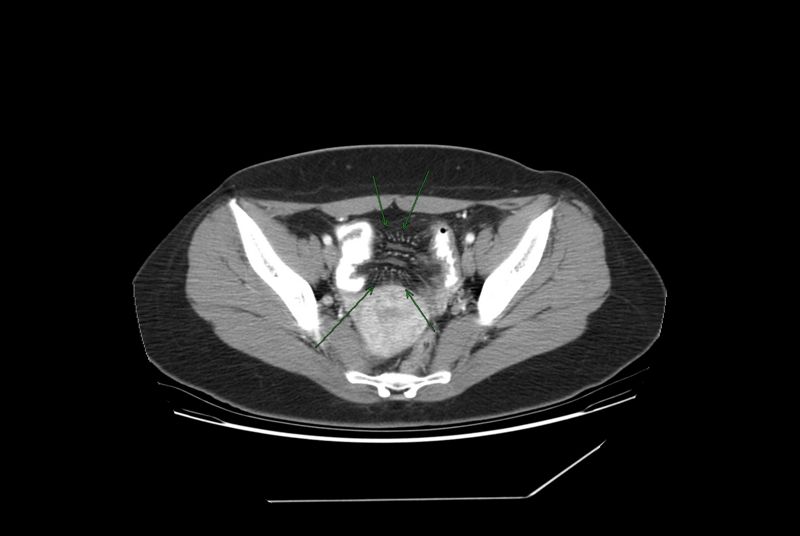
[[Image:Comb3.jpg|150px|thumb|left|Comb sign in Crohn's disease]] | [[Image:Comb3.jpg|150px|thumb|left|Comb sign in Crohn's disease]] | ||
==References== | ==References== | ||
Revision as of 15:52, 3 June 2013
|
Crohn's disease |
|
Diagnosis |
|---|
|
Treatment |
|
Case Studies |
|
Crohn's disease CT On the Web |
|
American Roentgen Ray Society Images of Crohn's disease CT |
Editor-In-Chief: C. Michael Gibson, M.S., M.D. [1]
CT
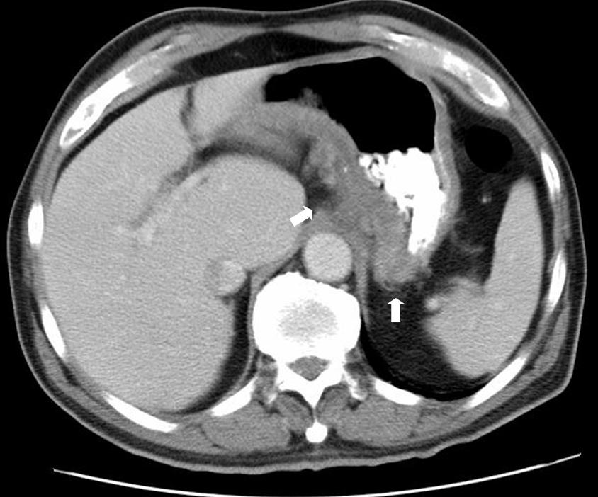
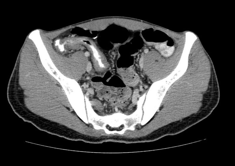
CT and MRI scans are useful for evaluating the small bowel with enteroclysis protocols.[1]They are additionally useful for looking for intra-abdominal complications of Crohn's disease such as abscesses, small bowel obstruction, or fistulae.[2] Magnetic resonance imaging (MRI) is another option for imaging the small bowel as well as looking for complications, though it is more expensive and less readily available.[3]


References
- ↑ Rajesh, A. (2006). "Multislice CT enteroclysis: technique and clinical applications". Clinical Radiology. 61 (1): 31–9. doi:10.1016/j.crad.2005.08.006. PMID 16356814. Unknown parameter
|coauthors=ignored (help); Unknown parameter|month=ignored (help) - ↑ Zissin, Rivka (2005). "Computed Tomographic Findings of Abdominal Complications of Crohn's Disease—Pictorial Essay" (PDF). Canadian Association of Radiologists Journal. 56 (1): 25–35. PMID 15835588. Retrieved 2006-07-02. Unknown parameter
|month=ignored (help); Unknown parameter|coauthors=ignored (help) - ↑ MacKalski, B. A. (2005). "New diagnostic imaging tools for inflammatory bowel disease". Gut. 55 (5): 733–41. doi:10.1136/gut.2005.076612. PMID 16609136. Unknown parameter
|coauthors=ignored (help); Unknown parameter|month=ignored (help)