Salmonella
|
Salmonellosis Microchapters |
|
Diagnosis |
|---|
|
Treatment |
|
Case Studies |
|
Salmonella On the Web |
|
American Roentgen Ray Society Images of Salmonella |
Editor-In-Chief: C. Michael Gibson, M.S., M.D. [1]; Associate Editor(s)-in-Chief: João André Alves Silva, M.D. [2] Jolanta Marszalek, M.D. [3]
Overview
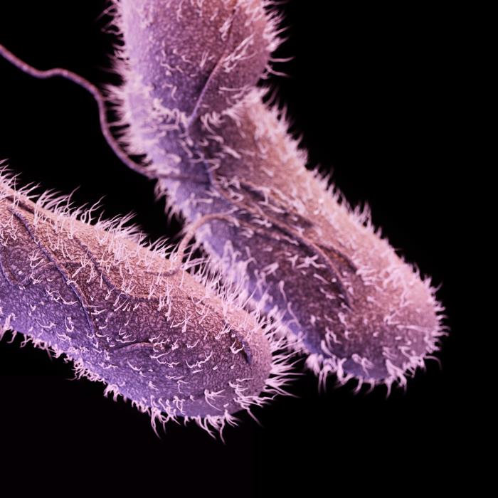
Salmonella is a genus of rod-shaped Gram-negative enterobacteria.[1] Salmonella species are motile and produce hydrogen sulfide.[2]
Taxonomy
Cellular organism; Bacteria; Proteobacteria; Gammaproteobacteria; Enterobacteriales; Enterobacteriaceae[3]
Biology
 |
 |
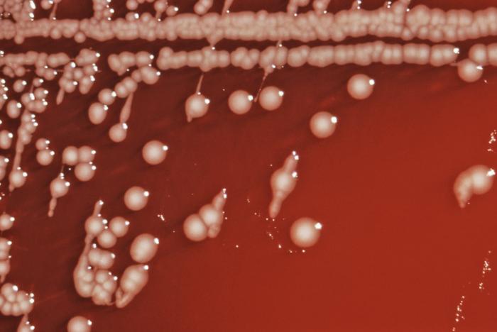
Salmonella is a Gram-negative bacterium, facultatively intracellular anaerobic, non-spore-forming bacilli. It measures 2 to 3 by 0.4 to 0.6 μm. Salmonellae do not ferment lactose, reduce nitrates, produce acid on glucose fermentation and are non producers of cytochrome oxidase.[5] Due to the presence of flagella, almost all salmonella are motile. 1% of the bacteria are able to ferment lactose, which may be responsible for its non-detection in media other than MacConkey agar.
For the isolation of salmonella in culture media, freshly passed stool are preferred. Common media for the growth of salmonella include: MacConkey agar, deoxycholate agar, and xylose-lysine-deoxycholate agar.[6]
When the sample has a low number of bacteria, special enrichment broths, such as the selenite-based enrichment broth, may be used to increase the number of bacteria.[6]
Salmonella isolates should be serogrouped with polyvalent antisera or in public health centers. Salmonella are serogrouped according to:[7]
- Capsular antigen
- Polysaccharide O antigens
- Flagellar antigens
Different salmonella serotypes may be distinguished according to the different metabolism of sugars.[8]
Infectious Cycle
Salmonella enterica enters the body through the mouth, by ingestion of contaminated food and water. For the bacteria to cause disease, an inoculum of about 50 000 bacteria is often required. Once in the intestine, the bacteria will first localize at the apical epithelium. Salmonella will then initiate bacterial mechanisms that allow host cell invasion, inducing inflammatory changes, such as:[9][10][11][12]
- Diffuse and focal infiltration of PMN
- Crypt abscess
- Necrosis of the epithelium
- Fluid secretion
- Edema
Different servers will have different preferable intestinal locations. An example is the enterocolitis at the terminal ileum, cecum, and proximal colon caused by serovar Typhimurium. Intestinal disease is marked by neutrophil migration to the intestinal epithelium. This recruitment is done by the secretion of interleukin-8, induced by Salmonella.[13]
Classification
Salmonella enteric may be subdivided As of December 7, 2005, there are two species within the genus: S. bongori (previously subspecies V) and S. enterica (formerly called S. choleraesuis), which is divided into six subspecies:[14][15]
- I—enterica
- II—salamae
- IIIa—arizonae
- IIIb—diarizonae
- IV—houtenae
- V—obsolete (now designated S. bongori)
- VI—indica
There are also numerous (over 2500) serovars within both species, which are found in a disparate variety of environments and which are associated with many different diseases. The vast majority of human isolates (>99.5%) are subspecies S. enterica. For the sake of simplicity, the CDC recommends that Salmonella species be referred to only by their genus and serovar, e.g.,
- Salmonella Typhi
instead of the more technically correct designation,
- Salmonella enterica subspecies enterica serovar Typhi.
Salmonella isolates are most commonly classified according to serology (Kauffman-White classification).[14] The main division is first by the somatic O antigen, then by flagellar H antigens. H antigens are further divided into phase 1 and phase 2. Both phase 1 and phase 2 H antigens are required for the full identification of an isolate but in practise, routine labs will leave this to Reference Laboratories.
Note that, with the exception of typhoid and paratyphoid, salmonellosis is not a blood-related infection, as is commonly believed.
Examples:
- Salmonella Enteritidis (1,9,12:g,m) - where the O antigens present are 1, 9 and 12; the H antigens are g and m.
- Salmonella Typhi (9,12,Vi:d:−) - where the O antigens are 9, 12,; the H antigen is d: The Vi antigen is associated with the bacterial capsule, which acts as a Virulence factor, hence its name.
In a clinical laboratory, only a small number of serovars are looked for (the remainder being rare or not clinically significant). The Health Protection Agency recommend testing for the following antigens routinely:
- O antigens: 2 4 6.7 8 9 and 3.10
- phase 1 H antigens: a b d E G i r Vi
- phase 2 H antigens: 1,2 1,5 1,6 1,7
Isolates that cannot be identified using this panel are sent to the reference laboratory for identification.
Tropism
Natural Reservoir
Related Chapters
- 1984 Rajneeshee bioterror attack
- List of foodborne illness outbreaks
- Food Testing Strips
References
- ↑ Ryan KJ, Ray CG (editors) (2004). Sherris Medical Microbiology (4th ed. ed.). McGraw Hill. ISBN 0-8385-8529-9.
- ↑ Giannella RA (1996). "Salmonella". In Baron S et al (eds.). Baron's Medical Microbiology (4th ed. ed.). Univ of Texas Medical Branch. ISBN 0-9631172-1-1.
- ↑ "Salmonella (Taxonomy)".
- ↑ Jump up to: 4.0 4.1 "Public Health Image Library (PHIL), Centers for Disease Control and Prevention".
- ↑ Mandell, Gerald (2010). Mandell, Douglas, and Bennett's principles and practice of infectious diseases. Philadelphia, PA: Churchill Livingstone/Elsevier. ISBN 0443068399.
- ↑ Jump up to: 6.0 6.1 Perez, J. M.; Cavalli, P.; Roure, C.; Renac, R.; Gille, Y.; Freydiere, A. M. (2003). "Comparison of Four Chromogenic Media and Hektoen Agar for Detection and Presumptive Identification of Salmonella Strains in Human Stools". Journal of Clinical Microbiology. 41 (3): 1130–1134. doi:10.1128/JCM.41.3.1130-1134.2003. ISSN 0095-1137.
- ↑ Murray, Patrick (2013). Medical microbiology. Philadelphia: Elsevier/Saunders. ISBN 0323086926.
- ↑ Mandell, Gerald (2010). Mandell, Douglas, and Bennett's principles and practice of infectious diseases. Philadelphia, PA: Churchill Livingstone/Elsevier. ISBN 0443068399.
- ↑ McGovern VJ, Slavutin LJ (1979). "Pathology of salmonella colitis". Am J Surg Pathol. 3 (6): 483–90. PMID 534385.
- ↑ Giannella RA, Formal SB, Dammin GJ, Collins H (1973). "Pathogenesis of salmonellosis. Studies of fluid secretion, mucosal invasion, and morphologic reaction in the rabbit ileum". J Clin Invest. 52 (2): 441–53. doi:10.1172/JCI107201. PMC 302274. PMID 4630603.
- ↑ Clarke RC, Gyles CL (1987). "Virulence of wild and mutant strains of Salmonella typhimurium in ligated intestinal segments of calves, pigs, and rabbits". Am J Vet Res. 48 (3): 504–10. PMID 3551701.
- ↑ Finlay BB, Heffron F, Falkow S (1989). "Epithelial cell surfaces induce Salmonella proteins required for bacterial adherence and invasion". Science. 243 (4893): 940–3. PMID 2919285.
- ↑ McCormick BA, Colgan SP, Delp-Archer C, Miller SI, Madara JL (1993). "Salmonella typhimurium attachment to human intestinal epithelial monolayers: transcellular signalling to subepithelial neutrophils". J Cell Biol. 123 (4): 895–907. PMC 2200157. PMID 8227148.
- ↑ Jump up to: 14.0 14.1 "The type species of the genus Salmonella Lignieres 1900 is Salmonella enterica (ex Kauffmann and Edwards 1952) Le Minor and Popoff 1987, with the type strain LT2T, and conservation of the epithet enterica in Salmonella enterica over all earlier epithets that may be applied to this species. Opinion 80". Int J Syst Evol Microbiol. 55 (Pt 1): 519–20. 2005. PMID 15653929.
- ↑ Tindall BJ; Grimont PAD, Garrity GM; Euzéby JP (2005). "Nomenclature and taxonomy of the genus Salmonella". Int J Syst Evol Microbiol. 55: 521&ndash, 524. PMID 15653930.
- CS1 maint: Extra text: authors list
- CS1 maint: Extra text
- CS1 maint: Extra text: editors list
- CS1 maint: Multiple names: authors list
- CS1 maint: PMC format
- Bacterial diseases
- Gram negative bacteria
- Enterobacteria
- Foodborne illnesses
- Zoonoses
- Disease
- Infectious disease
- Emergency medicine
- Intensive care medicine
- Rat carried diseases