No-reflow case 1: Difference between revisions
Hardik Patel (talk | contribs) Created page with "__NOTOC__ {{PCI}} {{CMG}}; {{AE}} {{HP}}, {{Sapan}} ==Abrupt Closure== Shown below is a pre-intervention angiogram with occlusion in the proximal part of SVG (saphenous..." |
Hardik Patel (talk | contribs) No edit summary |
||
| Line 8: | Line 8: | ||
Shown below is an angiogram with ongoing intervention on the occlusion described above. | Shown below is an angiogram with ongoing intervention on the occlusion described above. | ||
[[File:No-reflow-(13).gif|center|400px]] | [[File:No-reflow-(13).gif|center|400px]] | ||
Shown below is a post-intervention angiogram with acute reduction in [[blood flow]] ([[TIMI flow grade 1]]) in the absence of dissection, [[thrombus]], spasm, or high-grade residual stenosis in the [[SVG]] ([[saphenous vein graft]]) | Shown below is a post-intervention angiogram with acute reduction in [[blood flow]] ([[TIMI flow grade 1]]) in the absence of dissection, [[thrombus]], spasm, or high-grade residual stenosis in the [[SVG]] ([[saphenous vein graft]]) depicting no-reflow. | ||
[[File:No-reflow-(19).gif|center|400px]] | [[File:No-reflow-(19).gif|center|400px]] | ||
Shown below is a post-intervention angiogram with a complete restoration of [[blood flow]] ([[TIMI flow grade 3]]) after transient no-reflow as depicted above. | Shown below is a post-intervention angiogram with a complete restoration of [[blood flow]] ([[TIMI flow grade 3]]) after transient no-reflow as depicted above. | ||
Revision as of 18:15, 20 September 2013
|
Percutaneous coronary intervention Microchapters |
|
PCI Complications |
|---|
|
PCI in Specific Patients |
|
PCI in Specific Lesion Types |
|
No-reflow case 1 On the Web |
|
American Roentgen Ray Society Images of No-reflow case 1 |
|
Directions to Hospitals Treating Percutaneous coronary intervention |
Editor-In-Chief: C. Michael Gibson, M.S., M.D. [1]; Associate Editor(s)-in-Chief: Hardik Patel, M.D., Sapan Patel M.B.B.S
Abrupt Closure
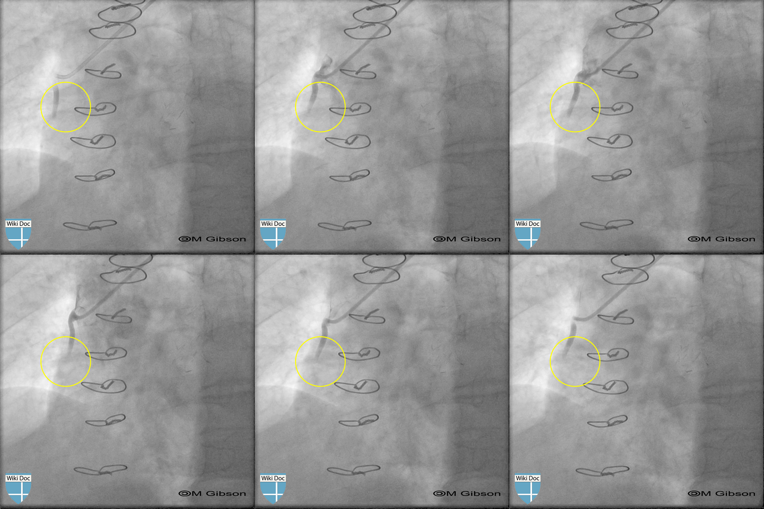
Shown below is a pre-intervention angiogram with occlusion in the proximal part of SVG (saphenous vein graft) to the RCA.

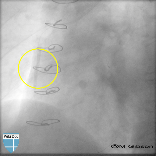
Shown below is an angiogram with ongoing intervention on the occlusion described above.

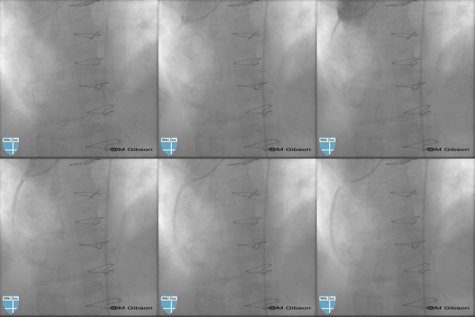
Shown below is a post-intervention angiogram with acute reduction in blood flow (TIMI flow grade 1) in the absence of dissection, thrombus, spasm, or high-grade residual stenosis in the SVG (saphenous vein graft) depicting no-reflow.

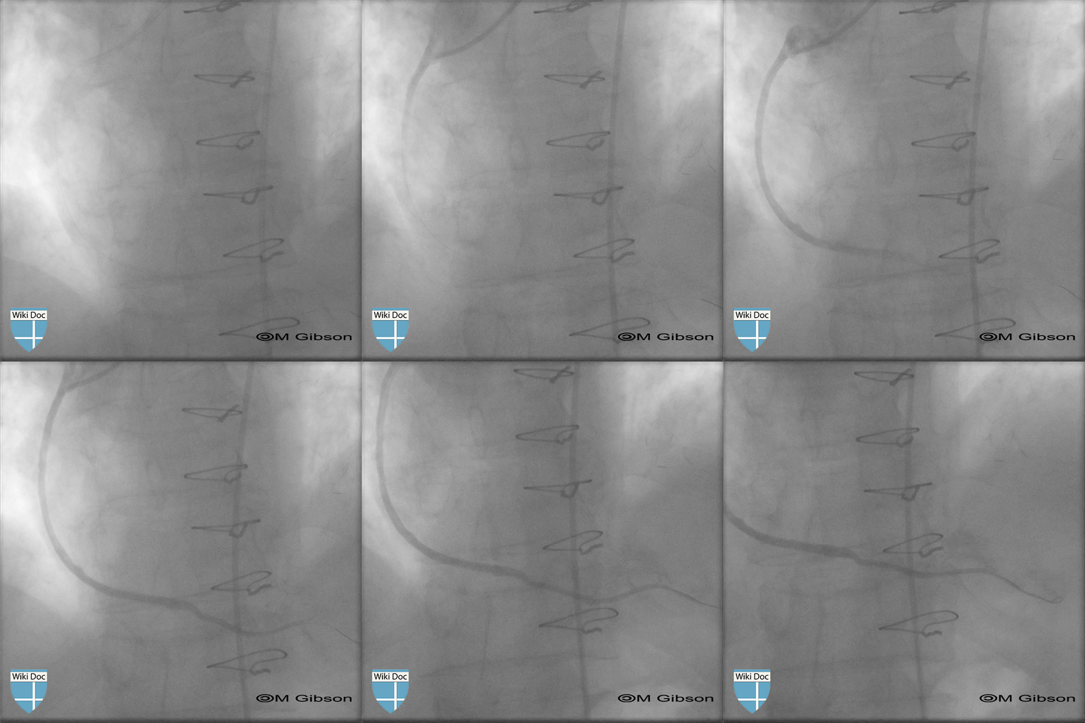
Shown below is a post-intervention angiogram with a complete restoration of blood flow (TIMI flow grade 3) after transient no-reflow as depicted above.
