Pneumonia CT: Difference between revisions
Jump to navigation
Jump to search
m (Bot: Removing from Primary care) |
|||
| (6 intermediate revisions by 3 users not shown) | |||
| Line 1: | Line 1: | ||
__NOTOC__ | __NOTOC__ | ||
{{Pneumonia}} | {{Pneumonia}} | ||
{{CMG}}; {{AE}} {{AL}} | {{CMG}}; {{AE}} {{HQ}}, {{AL}} | ||
==Overview== | ==Overview== | ||
A chest CT scan is not routinely done in patients with pneumonia, but is a diagnostic test that may be useful when a chest | A chest [[Computed tomography|CT scan]] is not routinely done in patients with pneumonia, but is a [[diagnostic test]] that may be useful when a [[Chest X-ray|chest x-ray]] is not conclusive. [[Computed tomography|CT]] findings may include lobar [[Consolidation (medicine)|consolidation]], ground-glass opacities, [[pleural effusion]], [[lymphadenopathy]], and tree-in-bud appereance. | ||
==CT== | ==CT== | ||
*A chest CT could be useful when a chest | *A chest [[Computed tomography|CT]] could be useful when a [[Chest X-ray|chest x-ray]] has inconclusive signs of pneumonia but the clinical manifestation suggest pneumonia. | ||
*CT findings in pneumonia include:<ref name="Ichikado2014">{{cite journal|last1=Ichikado|first1=Kazuya|title=High-Resolution Computed Tomography Findings of Acute Respiratory Distress Syndrome, Acute Interstitial Pneumonia, and Acute Exacerbation of Idiopathic Pulmonary Fibrosis|journal=Seminars in Ultrasound, CT and MRI|volume=35|issue=1|year=2014|pages=39–46|issn=08872171|doi=10.1053/j.sult.2013.10.007}}</ref> | *[[Computed tomography|CT]] findings in pneumonia include:<ref name="Ichikado2014">{{cite journal|last1=Ichikado|first1=Kazuya|title=High-Resolution Computed Tomography Findings of Acute Respiratory Distress Syndrome, Acute Interstitial Pneumonia, and Acute Exacerbation of Idiopathic Pulmonary Fibrosis|journal=Seminars in Ultrasound, CT and MRI|volume=35|issue=1|year=2014|pages=39–46|issn=08872171|doi=10.1053/j.sult.2013.10.007}}</ref> | ||
**Airspace [[Consolidation (medicine)|consolidation]] | |||
**Ground-glass opacities | |||
**[[Pleural effusion]] | |||
**Hilar and/or mediastinal [[lymphadenopathy]] | |||
**[[Bronchiectasis]] | |||
**Tree-in-bud appereance | |||
*A chest [[Computed tomography|CT]] can also help to assess reasons for therapy failure and complications, such as [[lung]] [[abscess]], and [[Pleural effusion|pleural effusions]]. | |||
{| | {| | ||
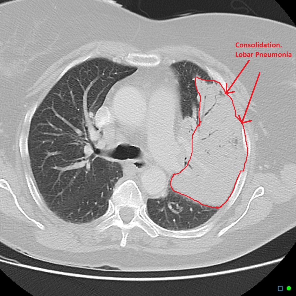
|[[ | |[[File:Lobar-pneumonia-ct-findings.jpg|300px|thumb|left| Areas of consolidation [https://radiopaedia.org/cases/lobar-pneumonia-ct-findings Source: Case courtesy of Dr Chris O'Donnell, Radiopaedia.org, rID: 32998]]] | ||
|} | |} | ||
====Comparison Between CT Findings in Viral and Bacterial Pneumonia==== | ====Comparison Between CT Findings in Viral and Bacterial Pneumonia==== | ||
{| style="border: 0px; font-size: 85%; margin: 3px; width:400px;" align=center | {| style="border: 0px; font-size: 85%; margin: 3px; width:400px;" align="center" | ||
|valign=top| | | valign="top" | | ||
|+ | |+ | ||
! style="background: #4479BA; color:#FFF; width: 200px;" | CT Finding | ! style="background: #4479BA; color:#FFF; width: 200px;" | CT Finding | ||
| Line 34: | Line 33: | ||
| style="padding: 5px 5px; background: #F5F5F5;text-align:center" | 33% | | style="padding: 5px 5px; background: #F5F5F5;text-align:center" | 33% | ||
|- | |- | ||
| style="padding: 5px 5px; background: #DCDCDC;font-weight: bold" | Focal Consolidation | | style="padding: 5px 5px; background: #DCDCDC;font-weight: bold" | Focal [[Consolidation (medicine)|Consolidation]] | ||
| style="padding: 5px 5px; background: #F5F5F5;text-align:center" | 9% | | style="padding: 5px 5px; background: #F5F5F5;text-align:center" | 9% | ||
| style="padding: 5px 5px; background: #F5F5F5;text-align:center" | 6% | | style="padding: 5px 5px; background: #F5F5F5;text-align:center" | 6% | ||
|- | |- | ||
| style="padding: 5px 5px; background: #DCDCDC;font-weight: bold" | Pleural Effusion | | style="padding: 5px 5px; background: #DCDCDC;font-weight: bold" | [[Pleural effusion|Pleural Effusion]] | ||
| style="padding: 5px 5px; background: #F5F5F5;text-align:center" | 41% | | style="padding: 5px 5px; background: #F5F5F5;text-align:center" | 41% | ||
| style="padding: 5px 5px; background: #F5F5F5;text-align:center" | 22% | | style="padding: 5px 5px; background: #F5F5F5;text-align:center" | 22% | ||
| Line 54: | Line 53: | ||
| style="padding: 5px 5px; background: #F5F5F5;text-align:center" | 31% | | style="padding: 5px 5px; background: #F5F5F5;text-align:center" | 31% | ||
|- | |- | ||
| style="padding: 5px 5px; background: #DCDCDC;font-weight: bold" | Multifocal Consolidation | | style="padding: 5px 5px; background: #DCDCDC;font-weight: bold" | Multifocal [[Consolidation (medicine)|Consolidation]] | ||
| style="padding: 5px 5px; background: #F5F5F5;text-align:center" | 36% | | style="padding: 5px 5px; background: #F5F5F5;text-align:center" | 36% | ||
| style="padding: 5px 5px; background: #F5F5F5;text-align:center" | 27% | | style="padding: 5px 5px; background: #F5F5F5;text-align:center" | 27% | ||
|- | |- | ||
| style="padding: 0px 5px; background: #F5F5F5;" | | colspan="3" style="padding: 0px 5px; background: #F5F5F5;" |<small>Adapted from American Journal of Roentgenology. 2011;197: 1088-1095<ref name="MillerMickus2011">{{cite journal|last1=Miller|first1=Wallace T.|last2=Mickus|first2=Timothy J.|last3=Barbosa|first3=Eduardo|last4=Mullin|first4=Christopher|last5=Van Deerlin|first5=Vivanna M.|last6=Shiley|first6=Kevin T.|title=CT of Viral Lower Respiratory Tract Infections in Adults: Comparison Among Viral Organisms and Between Viral and Bacterial Infections|journal=American Journal of Roentgenology|volume=197|issue=5|year=2011|pages=1088–1095|issn=0361-803X|doi=10.2214/AJR.11.6501}}</ref></small> | ||
|} | |} | ||
==References== | ==References== | ||
{{Reflist|2}} | {{Reflist|2}} | ||
{{WS}} | |||
{{WH}} | |||
[[Category:Pneumonia]] | [[Category:Pneumonia]] | ||
[[Category:Pulmonology]] | [[Category:Pulmonology]] | ||
[[Category:Emergency medicine]] | [[Category:Emergency medicine]] | ||
[[Category:Pediatrics]] | [[Category:Pediatrics]] | ||
[[Category:Disease]] | [[Category:Disease]] | ||
Latest revision as of 23:45, 29 July 2020
|
Pneumonia Microchapters |
|
Diagnosis |
|---|
|
Treatment |
|
Case Studies |
|
Pneumonia CT On the Web |
|
American Roentgen Ray Society Images of Pneumonia CT |
Editor-In-Chief: C. Michael Gibson, M.S., M.D. [1]; Associate Editor(s)-in-Chief: Hamid Qazi, MD, BSc [2], Alejandro Lemor, M.D. [3]
Overview
A chest CT scan is not routinely done in patients with pneumonia, but is a diagnostic test that may be useful when a chest x-ray is not conclusive. CT findings may include lobar consolidation, ground-glass opacities, pleural effusion, lymphadenopathy, and tree-in-bud appereance.
CT
- A chest CT could be useful when a chest x-ray has inconclusive signs of pneumonia but the clinical manifestation suggest pneumonia.
- CT findings in pneumonia include:[1]
- Airspace consolidation
- Ground-glass opacities
- Pleural effusion
- Hilar and/or mediastinal lymphadenopathy
- Bronchiectasis
- Tree-in-bud appereance
- A chest CT can also help to assess reasons for therapy failure and complications, such as lung abscess, and pleural effusions.
 |
Comparison Between CT Findings in Viral and Bacterial Pneumonia
| CT Finding | Bacterial | Viral |
|---|---|---|
| No findings | 9% | 33% |
| Focal Consolidation | 9% | 6% |
| Pleural Effusion | 41% | 22% |
| Ground-glass Opacity | 45% | 22% |
| Tree-in-bud Appereance | 14% | 31% |
| Bronchial Wall Thickening | 27% | 31% |
| Multifocal Consolidation | 36% | 27% |
| Adapted from American Journal of Roentgenology. 2011;197: 1088-1095[2] | ||
References
- ↑ Ichikado, Kazuya (2014). "High-Resolution Computed Tomography Findings of Acute Respiratory Distress Syndrome, Acute Interstitial Pneumonia, and Acute Exacerbation of Idiopathic Pulmonary Fibrosis". Seminars in Ultrasound, CT and MRI. 35 (1): 39–46. doi:10.1053/j.sult.2013.10.007. ISSN 0887-2171.
- ↑ Miller, Wallace T.; Mickus, Timothy J.; Barbosa, Eduardo; Mullin, Christopher; Van Deerlin, Vivanna M.; Shiley, Kevin T. (2011). "CT of Viral Lower Respiratory Tract Infections in Adults: Comparison Among Viral Organisms and Between Viral and Bacterial Infections". American Journal of Roentgenology. 197 (5): 1088–1095. doi:10.2214/AJR.11.6501. ISSN 0361-803X.