M-mode echo: principles and classic findings
Jump to navigation
Jump to search
Editor-In-Chief: C. Michael Gibson, M.S., M.D. [1]
Associate Editor-In-Chief: Cafer Zorkun, M.D., Ph.D. [2]
Overview
One-dimensional or M-mode echocardiography is one beam of ultrasound directed toward the heart.
Historical Perspectives
- One of the first modes available
- Excellent resolution
Keys to interpretation
- What structure is being imaged?
- Where is the cursor?
- Size
- Septum in HOCM
- Timing
- Tamponade - diastolic collapse
- Density
- Myxomatous leaflets, masses
Technology
A single crystal rapidly alternates between transmission and receiver modes with rapid updating result, rapidly moving structures (eg, valve leaflets) can be monitored for their characteristic motion very high temporal resolution
Specific Structures
Aortic Valve M-mode Analysis
- During systole do the aortic valve leaflets oppose the aorta?
- Are the leaflets thick and calcified (bright)?
- Possible to have normal appearance on m-mode if non-calcific
- Are leaflets open throughout systole - HOCM, low-output state
 |
Mitral Valve M-mode Analysis
- Anterior leaflet with E/A appearance of diastology
- Decreased EF slope in MS
- Scalloping of leaflet tip in end systole in prolapse
 |
Ventricular M-mode
- Ventricular Wall Thickness
- Ventricular Chamber Size
- Intraventricular Masses
 |
M Mode Pathologic States
M Mode in Aortic Stenosis
- Thickened, calcified leaflets
- Dense, persistent echoes replacing the normal motion patterns
- Post-stenotic aortic root dilation (> 35mm)
- LVH
- Eccentrically placed diastolic closure line (BAV)
 |
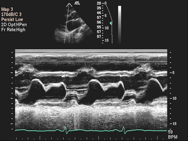
M Mode in Mitral Valve Prolapse
- Systolic bowing of the posterior mitral valve leaflet
 |
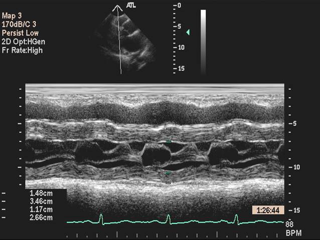
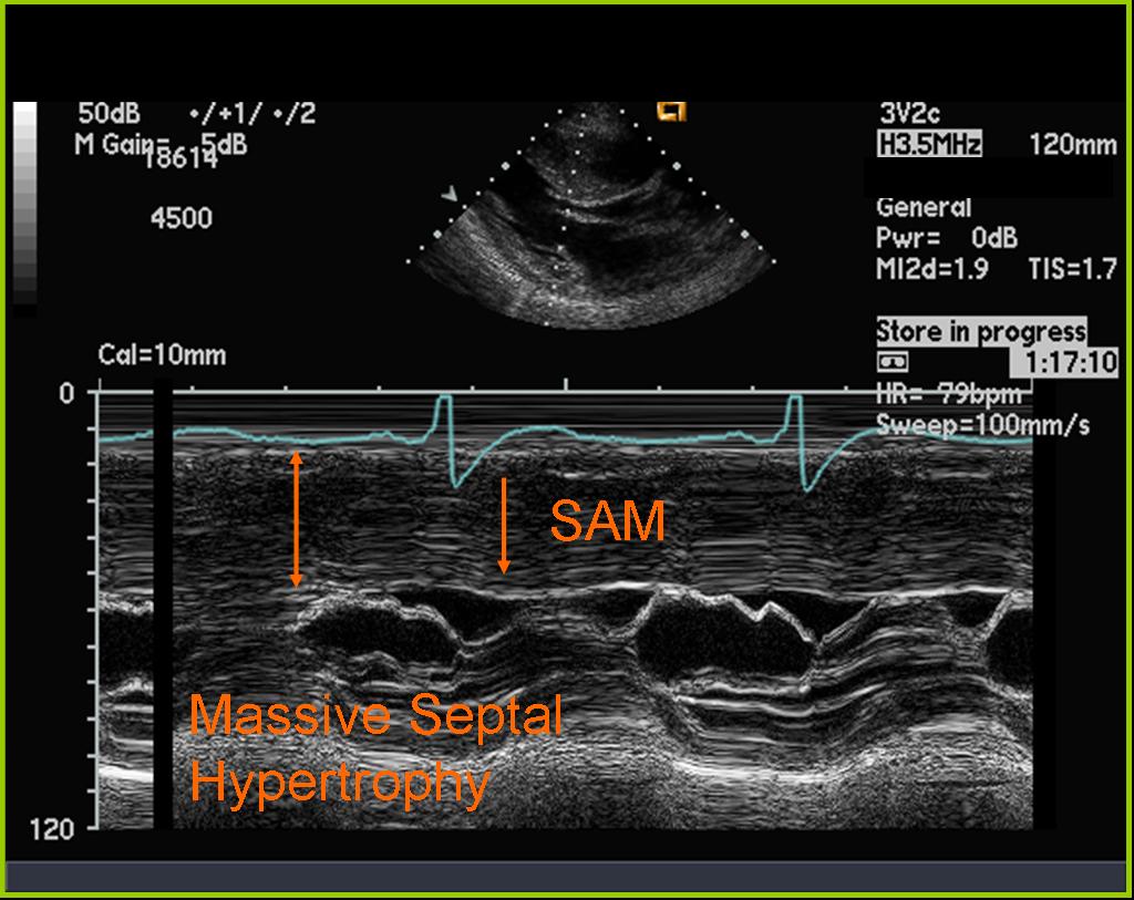
M Mode in Hypertrophic Cardiomyopathy
- Septal hypertrophy
- Systolic anterior motion (SAM) of the anterior mitral valve leaflet
- Mid-systolic (premature) closure of the aortic valve due to outflow track obstruction
 |
 |
 |
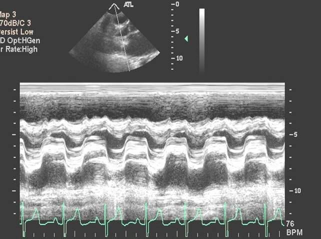
M Mode in Mitral Stenosis
- Leaflet tips bright (calcified) and thickened
- E/F slope decreased
 |
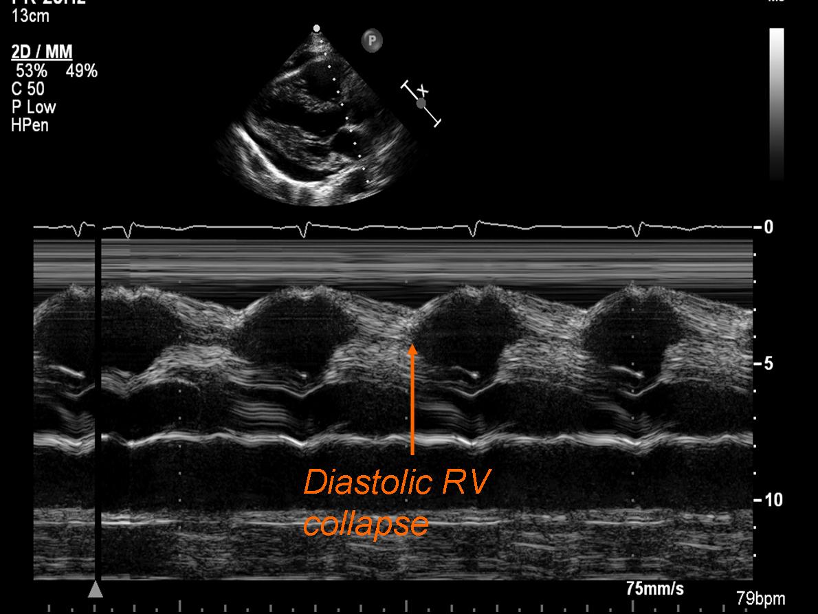
M Mode in Tamponade
- Diastolic collapse of the right ventricle
 |
 |
M Mode Aortic Regurgitation
- Increased duration between E and A peaks
- Fluttering of the anterior mitral valve leaflet due to AI jet turbulence
- Clinical setting to decide mechanism
 |
Other M Mode Findings
B Bump
- Elevated left ventricular end diastolic pressure
 |
M Mode in Endocarditis
- Thickened leaflets
- Good motion
- Mass
- Clinical scenario likely to give clue - dialysis patient, etc.
 |
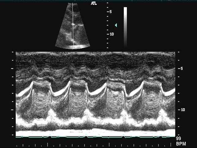
Left Atrial Myxoma
- Bright hyperdensity in mitral orifice throughout cardiac cycle
- Functional MS
 |
Pulmonary Hypertension
- M-mode of pulmonary valve - W appearance indicates elevated pulmonary pressures
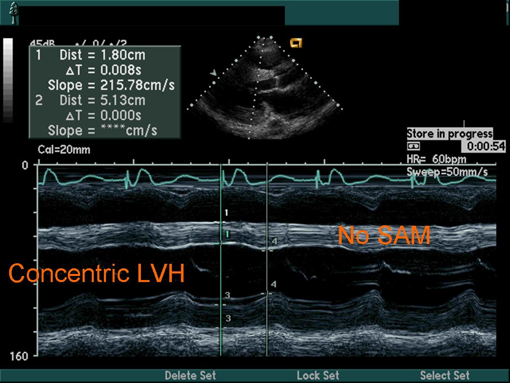
Hypertensive Heart Disease
 |
Mitral Stenosis
 |
Mitral Valve Prolapse
 |
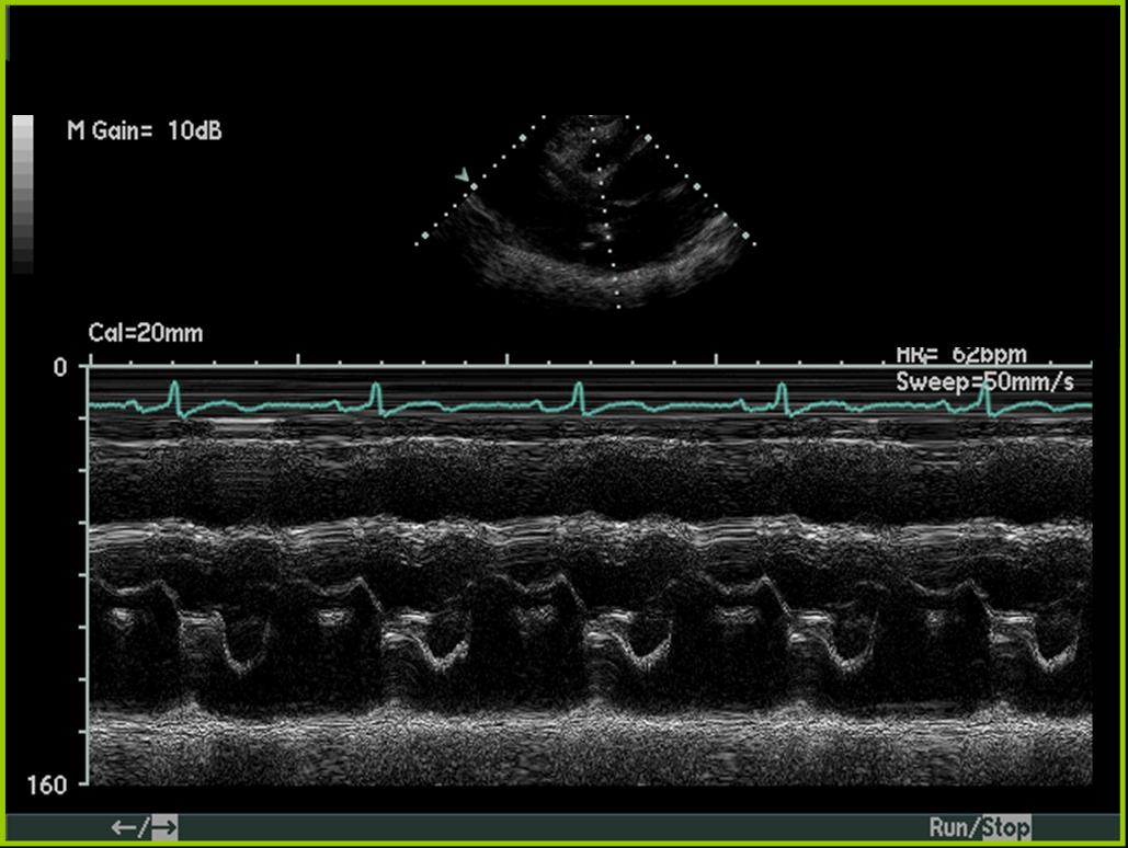
Mitral Regurgitation
 |