Chest X-ray

Template:Interventions infobox
|
WikiDoc Resources for Chest X-ray |
|
Articles |
|---|
|
Most recent articles on Chest X-ray Most cited articles on Chest X-ray |
|
Media |
|
Powerpoint slides on Chest X-ray |
|
Evidence Based Medicine |
|
Clinical Trials |
|
Ongoing Trials on Chest X-ray at Clinical Trials.gov Clinical Trials on Chest X-ray at Google
|
|
Guidelines / Policies / Govt |
|
US National Guidelines Clearinghouse on Chest X-ray
|
|
Books |
|
News |
|
Commentary |
|
Definitions |
|
Patient Resources / Community |
|
Patient resources on Chest X-ray Discussion groups on Chest X-ray Patient Handouts on Chest X-ray Directions to Hospitals Treating Chest X-ray Risk calculators and risk factors for Chest X-ray
|
|
Healthcare Provider Resources |
|
Causes & Risk Factors for Chest X-ray |
|
Continuing Medical Education (CME) |
|
International |
|
|
|
Business |
|
Experimental / Informatics |
Editor-In-Chief: C. Michael Gibson, M.S., M.D. [1]
Overview
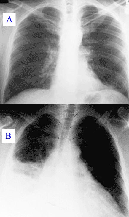
A chest X-ray, commonly abbreviated CXR, is a projection radiograph (X-ray), taken by a radiographer, of the thorax which is used to diagnose problems with that area.
Problems identified through chest x-rays
Examples of such problems include but are not limited to:
- Pneumothorax, sometimes tension pneumothorax (though this is usually diagnosed clinically because of its acute nature)
- Rib fracture
- Air space disease/consolidation (e.g. pneumonia)
- Interstitial lung disease (e.g. idiopathic pulmonary fibrosis (IPF), lung cancer, active tuberculosis)
- Cardiac silhouette enlargement - congestive heart failure, pericardial effusion, hypertrophic cardiomyopathies
- Pleural effusion
- Peritonitis
- Hiatal hernia
- Emphysema
- Pulmonary embolism (rarely) - usually CXR is normal
- Dissecting aortic aneurysm (due to trauma, advanced/untreated syphilis, connective tissue disorders)
Chest X-Rays are among the most common films taken, being diagnostic of so many important problems.
Features that are typically examined on a chest X-ray
Every doctor will have a different approach to examining chest X-rays. A commonly used mnemonic for what to look for on a chest X-ray is: It May Prove Quite Right (but) Stop And Be Certain How Lungs Appear:
- I = Identification (name, age, sex, indication for X-ray)
- M = Markers (differentiate left from right - diagnose dextrocardia)
- P = Position - the spinous process of T4 should be between the heads of the clavicle (if it isn't the body is rotated)
- Q = Quality - is the film penetrated properly. In a properly penetrated film the vertebral interspaces should be visible behind the central (cardiac) shadow
- R = Respiration - chest X-rays are typically done with full inspiration
- (but)
- S = Soft tissue - look for subcutaneous emphysema (suggestive of trauma), soft tissue swelling
- A = Abdomen - look for free abdominal air (suggests penetrating trauma, peritonitis, or recent surgery)
- B = Bone - look for fractures (these tend to be at the lateral aspects because of the mechanics - bending moment largest at lateral aspect)
- C = Central shadow (cardiac silhouette) - greater than 50% of lateral distance in frontal view at the diaphragm suggests cardiac enlargement (usually secondary to heart failure) or a pericardial effusion). A widened mediastinum may suggest aortic dissection.
- H = Hila (of the lungs) - can be affected in lung disease, malignant processes and infection (hilar lymphadenopathy).
- L = Lungs - for consolidation, interstitial lung disease (reticular, nodular or reticulonodular), honeycombing, miliary pattern, granulomas, lung masses
- A = Absent structures/Apices of the lung (for pneumothorax)
Another approach is to examine first any major abnormality, and then "review areas":
- the apices,
- the hila,
- behind the heart (it must be remembered that lung can be seen through the heart),
- the cardiophrenic angles,
- the costophrenic angles,
- beneath the diaphragm, and then
- bone and soft tissues.
A third approach is:
- Inspection of chest
- Centralized - Check whether the Xray is centralized. In a well centralized film the clavicle is equidistant from the spines of the vertebral body. It is important when we are commenting on the comparative radiolucency of the lung field, cardiomegaly and mediastenal shift.
- Compare the lung field on upper, middle, and lower zone to check for radiolucency on both side of lungs at equal levels.
- Check the apical area (just behind the clavicle) for the changes due to tuberculosis.
- Check for any cervical rib in cases of bony deformities
- Check for cardiophrenic and costophrenic angles. These are sharp and curved in healthy individuals. The right diaphragm is .5 - 1.5 cm higher than the left diaphragm.
- Check the hila for any hilar lymphadenopathy. The left hilum is slightly higher than the right
- Trace trachea up to the carina to check for any deviation.
- Inspection of the cardiac silhouette
- Check the cardiac borders. Left cardiac border is formed by aorta, pulmonary conus, left atrium, and right atrium from top to bottom. The right border is formed by superior vena cava and right atrium
- Measure the cardio-thoracic ratio to rule out any cardiomegaly. To measure the cardiothoracic ratio draw a vertical line through the spine of the vertebrae. Draw lines perpendicular from this line to the maximum width of the left (say for e.g. a) and right heart borders (say for e.g. b). The summation of a and b should be less than half of the maximum transverse diameter of the chest (say for e.g. c). Thus a+b = or < C/2
- Inspection of bones
- Check ribs for crowding, spreading and bony lesions
- Check clavicle, scapula, and shoulder joints
- Inspection of soft tissue
- Check chest muscles, subcutaneous tissue, and breast in females
Views
Typical views
- Frontal (view)
- PA (posterior-anterior)
- AP (anterior-posterior) - these are typically done in the ICU
- Lateral (view)
The most common view is the PA (posterior-anterior) and is frequently done with a left lateral view (so one can identify the location of abnormalities in 3-D space). PA views are generally preferred to AP views (which are often done with mobile/portable X-ray equipment), but much less convenient in the ICU setting or when a patient cannot otherwise leave their bed. PA views are preferred because the central shadow is better defined, the magnification of the heart is reduced, and less of the lungs obscured by the heart/pericardial sac.
Additional views
- Decubitus - useful for differentiating pleural effusions from consolidation (e.g. pneumonia). In effusions, the fluid layers out (by comparison to an up-right view, when it often accumulates in the costophrenic angles).
- Lordotic view - used to visualize the apex of the lung, to pick-up abnormalities such as a Pancoast tumour.
- Expiratory view - helpful for the diagnosis of pneumothorax
- Obliques
Chest X Ray findings in some common lung diseases
Tuberculosis chest x ray
Abnormalities
Nodule
A nodule is a discrete opacity in the lung which may be caused by:
- Neoplasm: benign or malignant
- Granuloma: tuberculosis
- Infection: round pneumonia
- Vascular: infarction, varix, Wegener's granulomatosis, rheumatoid arthritis
There are a number of features that are helpful in suggesting the diagnosis:
- rate of growth
- Doubling time of less than one month: sarcoma/infection/infarction/vascular
- Doubling time of six to 18 months: benign tumour/malignant granuloma
- Doubling time of more than 24 months: benign nodule
- calcification
- margin
- smooth
- lobulated
- presence of a corona radiata
- shape
- site
If the nodules are multiple, the differential is then smaller:
- infection: tuberculosis, fungal infection, septic emboli
- neoplasm: e.g., metastases, lymphoma, harmatoma
- sarcoidosis
- alveolitis
- auto-immune disease: e.g., Wegener's granulomatosis, rheumatoid arthritis
- inhalation (e.g., pneumoconiosis)
Cavities
A cavity is a walled hollow structure within the lungs. Diagnosis is aided by noting:
- wall thickness
- wall outline
- changes in the surrounding lung
The causes include:
- cancer (usually malignant)
- infarction (usually from a pulmonary embolus)
- infection: e.g., Staphylococcus aureus, tuberculosis, Gram negative bacteria (especially Klebsiella pneumoniae), and anaerobic bacteria.
Pleural abnormalities
Fluid in space between the lung and the chest wall is termed a pleural effusion. There needs to be at least 75ml of pleural fluid in order to blunt the costophrenic angle on the lateral chest X-ray, and 200ml on the posteroanterior chest X-ray. On a lateral decubitus, amounts as small as 5ml of fluid are possible. Pleural effusions typically have a meniscus visible on an erect chest X-ray, but loculated effusions (as occur with an empyema) may have a lenticular shape (the fluid making an obtuse angle with the chest wall).
Pleural thickening may cause blunting of the costophrenic angle, but is distinguished from pleural fluid by the fact that is occurs as a linear shadow ascending vertically and clinging to the ribs.
Diffuse shadowing
The differential for diffuse shadowing is very broad and can defeat even the most experienced radiologist. It is seldom possible to reach a diagnosis on the basis of the chest X-ray alone: high-resolution CT of the chest is usually required and sometimes a lung biopsy. The following features should be noted:
- type of shadowing (lines, dots or rings)
- reticular (crisscrossing lines)
- nodular (lots of small dots)
- rings or cysts
- ground glass
- consolidation (diffuse opacity with air bronchograms)
- location (where is the lesion worst?)
- upper zone (e.g., sarcoid, tuberculosis, silicosis/pneumoconiosis, ankylosing spondylitis, Langerhans cell histiocytosis)
- lower zone (e.g., cryptogenic fibrosing alveolitis, connective tissue disease, asbestosis, drug reactions)
- central (e.g., pulmonary oedema, alveolar proteinosis, lymphoma, Kaposi's sarcoma, PCP)
- peripheral (e.g., cryptogenic fibrosing alveolitis, connective tissue disease,chronic eosinophilic pneumonia, bronchiolitis obliterans organizing pneumonia)
- lung volume
- increased (e.g., Langerhans cell histiocytosis, lymphangioleiomyomatosis, cystic fibrosis, allergic bronchopulmonary aspergillosis)
- decreased (e.g., fibrotic lung disease, chronic sarcoidosis, chronic extrinsic allergic alveolitis)
Pleural effusions may occur with cancer, sarcoid, connective tissue diseases and lymphangioleiomyomatosis. The presence of a pleural effusion argues against pneumocystis pneumonia.
- Reticular (linear) pattern
- (sometimes called "reticulonodular" because of the appearance of nodules at the intesection of the lines, even though there are no true nodule present)
- cryptogenic fibrosing alveolitis
- connective tissue disease
- sarcoidosis
- radiation fibrosis
- asbestosis
- lymphangitis carcinomatosis
- Nodular pattern
- Cystic
-
- cryptogenic fibrosing alveolitis (late stage "honeycomb lung")
- cystic bronchiectasis
- Langerhans cell histocytosis
- lymphangioleiomyomatosis
- Consolidation
-
- Alveolar haemorrhage
- Alveolar cell carcinoma
- vasculitis
- chronic eosinophilic pneumonia
Limitations
It must be remembered that while the chest X-ray is a cheap and safe method of investigating diseases of the chest, there are a number of serious chest conditions that may be associated with a normal chest X-ray and other means of assessment may be necessary to make the diagnosis:
- Asthma
- Chronic obstructive pulmonary disease
- Pneumocystis jiroveci pneumonia (PCP)
- Pulmonary embolism
- Smoke inhalation
- Foreign body inhalation
Signs
Silhouette Sign
- The silhouette sign is especially helpful in localizing lung lesions. (e.g., loss of right heart border in right middle lobe pneumonia),[1]
Air Bronchogram Sign
- The air bronchogram sign, where branching radiolucent columns of air corresponding to bronchi is seen, usually indicates air-space (alveolar) disease, as from blood, pus, mucus, cells, protein surrounding the air bronchograms. This is seen in Respiratory distress syndrome[1]
Bean Sign
- The "bean" sign first described by Professor Keval Pandya is the appearance of a sharply circumscribed bean shaped nodule on chest X-ray which has a high sensitivity and specificity (92% and 88%) for the presence of miliary TB.
References
- ↑ Jump up to: 1.0 1.1 Chest X-Ray, OB-GYN 101: Introductory Obstetrics & Gynecology. © 2003, 2004, 2005, 2008 Medical Education Division, Brookside Associates, Ltd. Retrieved 9 February 2010.